POLARIZED LIGHT MICROSCOPY IN CONSERVATION: A PERSONAL PERSPECTIVEWALTER C. McCRONE
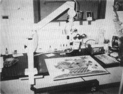
3 INSTRUMENTATIONOf the wide array of microscopes available today, those most useful to the conservator are the stereomicroscope and the PLM. If one has to economize, the PLM in combination with a stereomicroscope (both for less than $10,000) are the best choices. These two microscopes are adequate for inspecting, sampling, microchemical analysis, hot stage microscopy, as well as crystallographic characterization. Perhaps the most generally useful (and used) microscope is the stereomicroscope, called by biologists a dissecting microscope. It is essential for sampling such materials as paint layers, patinas, corrosion products, paper, inks, textiles, and metals. It resides immediately adjacent to the PLM in most laboratories. Although limited in magnification and resolving power (about 150x and 2 μm), it provides an erect (rather than a reversed left to right) stereo image. It is very useful for the examination and sampling of leather, ropes, paintings, inks, stains, insects, and biological damage, as well as for the examination of brush marks on, or cross sections of, paint layers. The stereomicroscope is not generally used for determining chemical composition of such materials, although many such objects in the 0.05–1 mm size range are recognized by shape, size, and color. The 2–3 inch working distance below the stereomicroscope objectives allows plenty of room for dissecting needles and probes. The conservator's stereomicroscope might well be on an extension arm to make possible the examination and sampling of paintings (fig. 5).
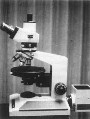
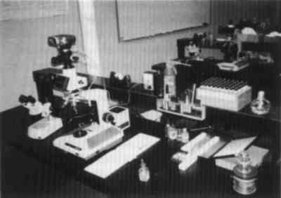
The conservator's PLM might also have an inclined binocular head for easier use (fig. 6), and a trinocular head will facilitate photomicrography or video microscopy. Not necessary are an automatic exposure camera, a heavy stand, confocal illumination, phase contrast, Nomarski, or Hoffman contrast accessories. A simple 35 mm or 4 � 5 Polaroid camera with an accessory exposure meter completes the major equipment needs of the conservation microscopist. A few books, reagents, solvents, microtools, refractive index liquids, slides, and coverslips (fig. 7) complete the basic microscopy laboratory.
If funds are available, two other very useful instruments are the SEM/EDS and the FTIR/microscope. If such instrumentation is unavailable internally, samples can be sent to a commercial laboratory. SEM/EDS is a method for elemental analysis of subnanogram samples suitable for the detection of most of the 104 chemical elements (at least sodium and above in the periodic table), but it is limited since it yields only an elemental analysis. Many combinations of pigments would yield similar EDS spectra, especially since the elements below sodium in the periodic table are “invisible” to EDS when using a detector with a beryllium window. For example, the lead oxides PbO and Pb3O4 would yield identical spectra—identical, as well, to the spectra of lead white and lead carbonate. SEM/EDS is most useful for differentiating between pigment look-alikes (e.g., ultramarine and smalt, hematite and vermilion), and an experienced PLM analyst is often relieved to have confirmation by SEM/EDS of such look-alikes in a given sample. Wavelength dispersive x-ray analysis by the far more expensive analytical transmission electron microscope or electron microprobe has a higher energy resolution and lower detection limits than EDS. The ion microscope (microprobe) covers all of the periodic table with subpicogram sensitivity. These latter instruments are quantitatively quite accurate. FTIR/microscopy is also an excellent supplement to PLM for the identification of pigments (Low and Baer 1977). It has the added advantage that it also characterizes media, varnishes, adhesives, and coatings. The FTIR/microscope yields an infrared absorption pattern for subnanogram samples. Like the SEM/EDS, however, it identifies parts of the molecules but, often, not how the parts are assembled. If the sample is a pure substance, pure only in the sense of a wax, varnish, paint medium, etc., the FTIR/microscope may well identify the sample as such and often even whether the wax is beeswax, camauba, etc. Other microanalytical instruments are only very occasionally useful to the conservator. While a few other analytical tools may identify some subnanogram samples, no instrument or technique other than PLM identifies all such particles, whether biological (e.g., species of pollen, wood, animal hair, etc.), crystalline or noncrystalline, organic or inorganic. Few analysts are able to isolate, clean, and mount a single subnanogram particle for x-ray diffraction analysis, SEM/EDS, or FTIR/microscopy. The PLM analyst does this routinely; still, it is not always necessary to separate individual particles from a matrix, as they are characterized and identified even in mixtures. Particles are visually separate, one from the other, in the microscope field of view. |