POLARIZED LIGHT MICROSCOPY IN CONSERVATION: A PERSONAL PERSPECTIVEWALTER C. McCRONE
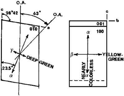
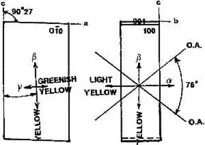
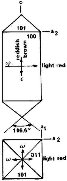
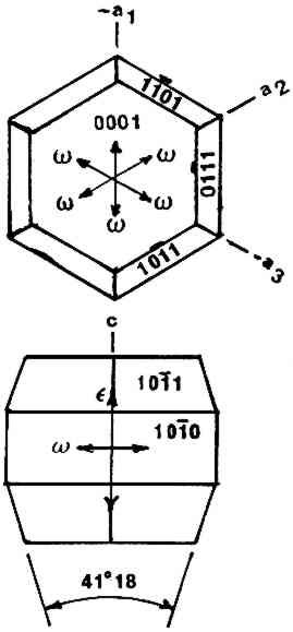
2 USES OF MICROSCOPYMicroscopy is defined as the application of any enlarged-image process enabling visualization of objects nominally invisible to the unaided human eye. Individuals using the instruments capable of producing enlarged images of tiny objects are called microscopists. Those individuals, properly trained, are able to extend the observation and interpretation of large (macroscopic) objects down to subnanometer objects difficult to identify by any other means. The PLM, by producing enlarged images and by examining samples between polarizing filters, makes it possible to see, isolate, identify, measure, interpret, and evaluate microscopic objects (Chamot and Mason 1958; Hartshorne 1960). After microscopical enlargement, most tiny pigments, fibers, etc., are usually identified by a process all of us use thousands of times a day to recognize people, buildings, trees, books, cars, etc. Many microscopic substances enlarged 10-1000x are then recognized just as definitely, and just as rapidly, as we recognize macroscopic objects without magnification. There are look-alikes, however, that require even experienced microscopists to hesitate, consider the possibilities, and decide how to differentiate between them (e.g., bast fibers, starch grains, wood fibers, and some blue or yellow pigments). Then more definitive measurements must be made. Usually a few simple and rapid tests are all that is required to reduce the number of possibilities, most often, to one. These might include closer attention to shape characteristics, measurement of refractive index, melting point, difference between the refractive indices (birefringence), or orientation of the refractive indices relative to “crystallographic” directions in the particle or fiber. For example, corn and rice starch, otherwise identical, differ in size by a factor of 2 (10 μm and 5 μm diameters, respectively); alizarin has a refractive index higher than the standard refractive index liquid (nD = 1.66), while its look-alike, madder, is lower; whiting has one refractive index equal to nD = 1.66 and is rhomb-shaped, while other common carbonate minerals with identical shapes all have higher indices. Fortunately, each of the pigments, as well as other crystalline substances, have a unique set of physical measurements (McCrone et al. 1984) that are made by PLM to ensure identification. Figures 1–4 show some of the crystallographic data for four representative pigments. No other substance in the universe is likely to match any of these four sets of morphological and optical data.
The PLM, unlike most other microanalytical tools, is equally applicable to organic, inorganic, biological, crystalline, or noncrystalline unknowns. It does, however, have the major disadvantage that a great deal of training and experience are necessary for best use. One might consider that becoming a competent microscopist is equivalent to learning to play a musical instrument well. After an introduction to the methods of microscopical analysis through formal instruction,1 these methods are best learned by actual observation and practice. The courses available (e.g., microchemical analysis, fiber and hair analyses, polarized light microscopy for the conservator, etc.) only tell what can be done and how to perform the techniques for proper use of the microscope and characterization of substances of interest. The student must observe, practice, and learn to use those techniques “on the job.” PLM data are far more definitive than the data obtained by SEM/EDS (x-ray microanalysis with the scanning electron microscope) data or even FTIR/microscopy (Fourier transform infrared absorption spectroscopy with microscope attachment), two widely used micro-analytical instruments. PLM not only identifies any pigments but also different forms of, say, lead white, lead oxides, or iron oxides. Lead white, the carbonate PbCO3 or the basic carbonate 2PbCO3�Pb(OH)2, are easily differentiated. There are several lead oxide pigments, including red lead, massicot, and litharge; each is distinctive in microscopical characteristics. These two other recently developed microanalytical instruments are, however, excellent supplements to the use of PLM. |