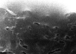
Sample at 60% RH; 15 sec.; 0.665MB; 1:75 compression
Click the image to download video.
This video documents an experiment on the effect of water on albumen photographs. The experiment was conducted using an environmental scanning electron microscope (ESEM) and is documented in a Messier & Vitale article, in the Library. The use of the ESEM has many advantages over standard SEM. The most obvious advantage is that the sample can be viewed real-time under controlled environmental conditions, such a the presence of water vapor. Experiments are not limited to static observations, or desiccated organics. Relative humidity and the presence of liquid water can be controlled by manipulation of the temperature and pressure within the sample chamber.
In this experiment, the albumen print sample was placed into a deeply grooved metal mount attached to a movable and temperature-controlled (Peltier effect) stage. The mount incorporated no adhesive or mechanical clamping. The sample was able to expand, contract and distort freely within the 0.05 mm slot. The overall length of the sample was 3 mm. This experiment was conducted at the ElectroScan Corporation during July, 1991. The sample can be seen to move around as it shrunk and curled; Trisha Rice McGrath did an excellent job of keeping the selected albumen segment in the field of view.
Three QuickTime video clips follow. Each segment is in real time (not time-lapsed), but has been edited to yield a small file size, which will be relatively quick to download. Narration was not included. The entire experiment took 23 minutes.
The first video segment shows the sample in cross-section a 1,000X magnification. A discrete layer of albumen is clearly visible. Two cracks in the albumen are also visible. The experiment was begun with the sample chamber at 60% RH, 3.9 Torr water vapor partial pressure, at 5 degrees C. The sample chamber has its water vapor partial pressure increased to just short of 100% RH.
Sample at 60% RH; 15 sec.; 0.665MB; 1:75 compressionClick the image to download video.
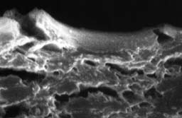
The next segment of the video shows the gradual immersion of the sample in water. The water vapor in the chamber condensed on the cooler surface of the metal sample holder. Immersion was achieved by increasing the vapor pressure to 7.8T at 5 degrees C. This segment ends with the sample totally immersed.
Immersing the Sample; 26 sec.; 0.484MB; 1:190 compressionClick the image to download video.
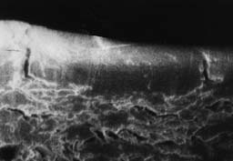
This last video segment shows the sample as it dries fully wet at the beginning. The first indication of drying is the opening is the opening of the paper pores, especially noticeable in the lower left of the screen. Drying was achieved approximately 2 minutes after decrease in water vapor pressure and warming of the stage started. The sample was brought to approximately 66% RH with the sample stage set at 5.7 degrees C and the sample chamber set at 4.7 Torr. Eventually, the sample was dried in equilibrium with 14% RH, in the chamber, at 4.2 Torr and 29 degrees C.
Drying the Sample; 52 sec.; 1 MB; 1:182 compressionClick the image to download video.
This experiment clearly shows the effect of the vigorous response of the albumen layer to water. The length of the center albumen sample began at 76 microns; it swelled 5% and then shrank to 91% of its original of its original length at 14% RH. It swelled in thickness 22% from 10.5 microns and shrank approximately the sample amount in response to wetting and drying. Another interesting results is the morphology of the paper is more open after wetting than before.
The ESEM provided a powerful tool to demonstrate the type and magnitude of change brought about by water and helped to focus a materials science investigation of albumen photographs. Images gained from the ESEM graphically illustrate the conclusions of the study that showed the weak albumen layer ruptures under the strain of its swelling and shrinkage in response to water and water vapor.
The video was captured in a miniDV dub from a consumer VCR recording (made directly from the ESEM), using a pro-sumer SVHS VCR. To achieve a small file size (350-1000 KB) from the 75-180 MB fcp.mov files, we used the Sorenson Video codex set "low," at 8 fps and 21-45Kb/s bit-rate, sized at 180 X 120 pixels (quarter original). Various amounts of compression were used to yield detail at important points in the QuickTime movies. Lower compression decreased pixelation.
We wish to acknowledge Trisha Rice McGrath and Edward Griffith III of the ElectroScan Corporation. A full report of this experiment, Cracking in Albumen Photographs: an ESEM Investigation, which appears in the Journal Microscopy Research and Technique, 1993, V.25, pages 374-383; who we also thank for allowing it to be presented here.
-- Tim Vitale and Paul Messier, 1993.