TECHNICAL STUDY OF ETHIOPIAN ICONS, NATIONAL MUSEUM OF AFRICAN ART, SMITHSONIAN INSTITUTIONErica E. James
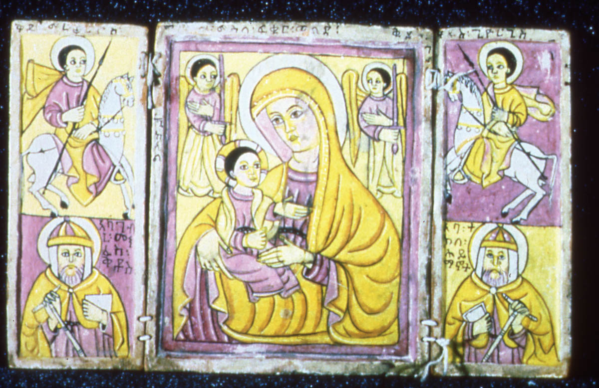
3 TECHNICAL STUDY: VISUAL EXAMINATIONThe first step in documentation was before-treatment photography with examination of the recto surface in normal and raking light and the verso surface in normal light. This photodocumentation was completed using tungsten lighting and Kodak Ektachrome color reversal film ASA 160. In addition, Plus-X Pan black-and-white print film ASA 125 was used to document the recto surface of each icon using normal light. Ultraviolet fluorescence examination was accomplished using tungsten lighting, Kodak Ektachrome color reversal film ASA 160, and a Kodak no. 2B Wratten Gelatin filter. Figure 2 illustrates the fluorescence typical of the icons. The purpose of the UV fluorescence examination was to ascertain the presence of varnish on the surface, to look for the presence of a pink fluorescence often associated with a madder lake paint, and to detect any retouching fluorescence. In visible light, the icons appeared to be unvarnished, and this finding was confirmed with ultraviolet fluorescence examination. One diptych (acc. no. 98-3-3) appeared in visible light to contain madder. Although ultraviolet light examination did not confirm this suspicion, madder was identified on this icon later through dispersed pigment analysis. Retouching was detected on acc. no. 73-24-1 along the lengthwise split in the center panel. Infrared reflectography (fig. 3) and x-ray radiography were also used to detect any losses and/or underdrawings in the paint layer and to discern variable densities of the pigment particles composing the paint layer, respectively. Infrared reflectography (film-based) was accomplished using tungsten lights, Kodak Ektachrome Professional Infrared EIR film (ASA 100/ Process E-6), and a Kodak no. 12 Wratten Gelatin filter. At the NMAfA, IR examination was conducted in Ektachrome slide format. The infrared technique detected only underdrawings that had already been seen during visible examination, thus providing no additional information concerning possible underdrawings not visible to the naked eye. Overall, however, the infrared technique did emphasize how linear and stylized the images in the icons are. Almost all the figures in the icons are outlined with black lines. During x-ray radiography, top views of all the icons were taken with Kodak ReadyPack x-ray film under the following conditions: 70 KV (kilovolts)/5 ma (milliamperes)/116cm TFD (total focal distance
In viewing the x-radiographs of the icons, the goal was to compare the relative densities of the paints on each icon to elucidate materials employed. The areas that were painted red appeared more radio-opaque than other areas due to the density of the red pigment. In acc. nos. 98-2-1, 94-21-1, 98-3-1, 98-3-2, and 73-24-1, the density of one pigment was predominant in the red areas of the paint layer. Cinnabar is mercuric sulfide (HgS) and is the only radio-opaque pigments typically used on the Ethiopian icons. Later analysis by polarized light microscopy did identify the pigment as cinnabar. The x-radiographs revealed no significant deviations in image composition or reconstruction from the final appearance of the icons. |
