IDENTIFICATION OF COLORANTS ON MAPS FROM THE EARLY COLONIAL PERIOD OF NEW SPAIN (MEXICO)MARY ELIZABETH HAUDE
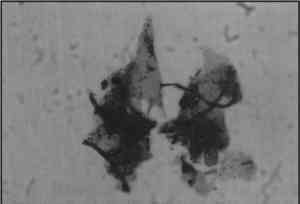
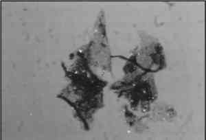
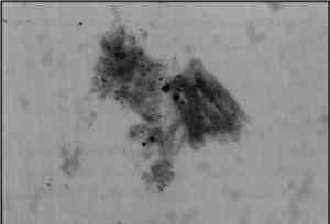
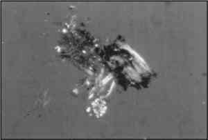
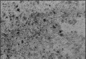
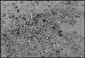
5 ANALYSIS OF COLORANT SAMPLESLaura Guti�rrez-Witt, head librarian for the Benson Latin American Collection, generously granted permission to remove small colorant samples from the six maps. Guti�rrez-Witt enthusiastically supported the pigment analysis because the Relaciones Geogr�ficas are among the most important artifacts at the Benson Latin American Collection and are continually used by scholars. While much is known as to why the RGs were made, she indicated that no one had researched how they were made; thus the pigment analysis would provide some insight into the types of paints and inks used on the maps. I sampled 22 colorants; the maps contain more than 22 colors, but the paints and inks are thinly applied in many areas. Sampling was therefore restricted to areas of heavy application. I was conservative in the sampling process because the paint layer consisted of washes directly applied to the paper substrate. Because losses in the paint layer could be easily detected, I refrained from removing large and numerous pigment samples. Under magnification, the samples As much as possible, I sampled colorants from painted areas showing both indigenous and European artistic influences. For instance, seven blue and blue-green paint samples were taken from European and Native pictorial elements on five of the maps. These seven paint samples were identified as Maya blue, an indigenous Mexican colorant. I identified 10 colorants from the 22 samples; many of the colorants could not be identified. Factors hindering positive identification included the extremely small size of the particles and the lack of sufficient standard samples with which to compare them. The budget and schedule for this study were limited. Optical microscopy was most available and therefore was chosen as the analytical method. Ideally, further methods would have been utilized to verify or invalidate the results obtained by microscopy. Microchemical analysis was considered but was difficult to perform because the samples were small and were mounted on microscope slides. However, a representative sample of the mounted pigments was sent to Robin Clark, head of the Department of Chemistry of University College, London, to be analyzed by Raman microscopy. Unfortunately, the results were inconclusive because the small sizes of the particles hindered positive identifications. The pigment samples were analyzed under 32x, 80x, and 128x by transmitted polarized light microscopy in uncrossed and partially crossed polars. I compared the samples to McCrone pigment standards and to the standards of Sara McElroy, painting conservator at UT-Austin's Archer M. Huntington Art Gallery, and identified several. Microscopically the particles range from 0.007 to 0.02 mm. Study of the refractive indices of the individual pigments was conducted at the Getty Conservation Institute with the help of David Scott, head of the Museum Services Scientific Program. This article will describe the individual colorants sampled from the maps. Table 2 (p. 266) summarizes the color of the paint, the identification of colorants, the optical characteristics of the colorants, and the maps from which they were sampled. TABLE 2 SUMMARY OF IDENTIFIED COLORANTS 5.1 RED COLORANTS5.1.1 CochinealThree red samples were removed from Ameca, Atlatlauca, and Cholula and identified as likely being cochineal. Microscopically these samples appear to be lake pigments where a clay base is dyed with an organic colorant. Ideally the organic component of the red samples would have been identified definitively by additional analytical methods (chemical, chromatography, or spectroscopy). According to Schweppe and Roosen-Runge (1986), the optical characteristics of cochineal include red particles that are translucent, granular, and isotropic. In uncrossed polars the red particles of the sample appear bright, cool red, translucent, and granular (fig. 6a, p. 253). In partially crossed polars the samples are isotropic, exhibiting the same physical properties in every direction as the microscope stage is rotated (fig. 6b, p. 253). The red particles are of a lower refractive index than the mounting medium.
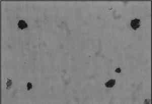
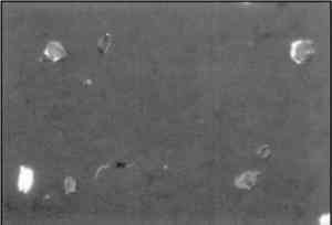
5.1.2 CinnabarA yellow-orange colorant was sampled from Meztitlan. Microscopically the sample consists of two distinct unidentified yellow particles and two distinct red particles. One red was identified as cinnabar. In uncrossed polars these particles are bright red, differ in size, and appear as fractured crystals; in partially crossed polars the particles appear 5.1.3 Red LeadThe other red particles in the yellow-orange colorant sample from Meztitlan were identified as red lead. In uncrossed polars the red particles are orange in color (fig. 7a, p. 253). In partially crossed polars these particles exhibit “blue-green interference colors” which Fitzhugh (1986) indicates is characteristic of red lead (fig. 7b, p. 253). The particles of red lead are of a higher refractive index than the mounting medium.
5.2 BLUE COLORANTS5.2.1 AzuriteA bright blue colorant sampled from Meztitlan microscopically consists of two distinct blue particles. One blue was identified as azurite and the other as Maya blue. In uncrossed polars the particles vary from dark to light blue and are slightly green. In partially crossed polars they are strongly birefracting, exhibiting different physical properties in different directions. (The blue particles blink as the microscope stage is rotated.) Gettens and Fitzhugh (1993) indicate that azurite is strongly birefracting and in uncrossed polars consists of coarse deep blue and finer pale blue particles with greenish undertones. The refractive index of the azurite particles is higher than that of the mounting medium. 5.2.2 IndigoA dark blue sample from Tehuantepec and a blue-green sample from Meztitlan were identified as likely being indigo. In uncrossed polars the particles of both samples are dark blue and uneven in size. In partially crossed polars the blue particles are isotropic. The indigo particles are slightly lower than the refractive index of Permount. Individual paticles of indigo are often difficult to resolve with optical microscopy and difficult to distinguish from Prussian blue (Schweppe 1997). Microchemical analysis of the two blue samples would provide more definitive results on their identity. However, I am certain that the aforementioned blue samples are indigo given the provenance of the RG maps and the abundance and documented use of indigo in 16th-century New Spain. 5.2.3 Maya BlueSeven green, blue, and blue-green colorants were sampled from Ameca, Atlatlauca, Ixtapalapa, Meztitlan, and Tehuantepec, and all were identified as Maya blue. In uncrossed polars the colorant samples are bright green-blue in color and translucent (fig. 8a, p. 253). In partially crossed polars the colorant samples are pleochroic, appearing blue in one direction and pink in another (fig. 8b, p. 253). (Pleochroism refers to the color change of particles in polarized light as the microscope stage is rotated.) Gettens and Stout (1966) describe Maya blue as blue in one direction and yellow in the other, but close microscopic examination of the Maya blue standard revealed a strong pleochroism from blue to pink in partially crossed polars. Given the blue to pink pleochroism of all seven colorant samples, the samples were identified as Maya blue. The Maya blue particles are in extremely low relief in Permount (n = 1.518–1.521); the refractive index of the clay base is close to 1.52, and the organic component is even lower.
5.3 GREEN COLORANTS5.3.1 Green EarthA yellow-xsgreen colorant sample from Meztitlan was identified as likely being green earth. Microscopically the sample contains green particles of various shades and a scattering of yellow clear, brown, and bright blue particles (fig. 9a, p. 257). The particles are pale, rounded, and translucent, with some exhibiting a grainy texture. In partially crossed polars the particles are moderately birefringent, or doubly refracting, blinking as the microscope stage is rotated (fig. 9b). Grissom (1986, 150) describes the optical characteristics of green earth as “particles of various shades of green intermixed with traces of yellow and brown earths” that are rounded, translucent, and low to moderately birefringent in polarized light. In addition, Gettens and Stout (1966) indicate that green earth consists of clear and bright blue particles. The green earth particles are of a higher refractive index than the mounting medium.
5.4 BROWN COLORANTS5.4.1 Raw UmberTwo brown colorant samples from Meztitlan and Tehuantepec were identified as raw umber. 5.4.2 Burnt UmberA third brown sample from Atlantlauca was identified as burnt umber. Microscopically this sample is similar to the raw umber samples, but it is more red. Also, the brown colorant sample and the burnt umber standard contain similar dark blue, almost purple, spots within the brown particles. The burnt umber particles are of a higher refractive index than the mounting medium. 5.5 BLACK COLORANTS5.5.1 Charcoal BlackOne black sample from Ixtapalapa was identified as charcoal black. Microscopically the black particles are elongated and splintery (figs. 10a, 10b, p. 257). Gettens and Stout (1966, 104) describe the optical characteristics of charcoal black as “small, opaque, elongated,
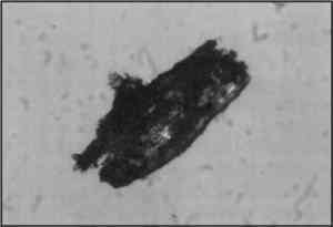
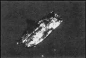
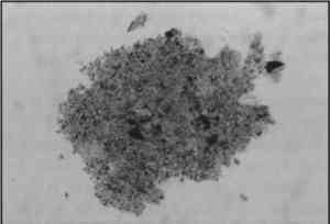
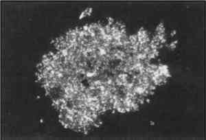
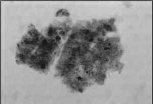
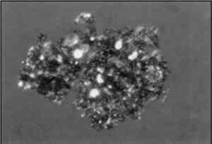
5.6 UNIDENTIFIED COLORANTSI was unable to identify four colorants from three of the maps. Three samples contain two distinct yellows, most likely organic. Figures 11a and b (p. 258) show photomicrographs of one of these unidentified yellows. The fourth sample is a light green dyed pigment, also likely organic (figs. 12a and b, p. 258). Table 3 (p. 267) provides a summary of the optical characteristics of the unidentified pigment samples. These samples were examined by UV-fluorescence microscopy by Narayan Khandekar, associate scientist at the Getty Conservation Institute.
TABLE 3 SUMMARY OF UNIDENTIFIED COLORANTS 5.7 RESULTSIn my opinion it is likely that the ten identified and four unidentified colorants on the six RG maps are of indigenous Mexican origin. Many of these pigments and dyes are mentioned in the historical literature; others have been identified on various pre- and postconquest artifacts. In addition, many inorganic pigments occur naturally throughout Mexico. While I cannot prove that these pigments were manufactured in 16th-century New Spain, I have found no evidence thus far of their importation into the New World. My literature survey also revealed the extensive knowledge and expertise that indigenous Mexican peoples had for making a wide variety of organic and inorganic colorants. I was not surprised to find cochineal, indigo, and charcoal black on the maps. These three colorants are well documented in the 16th-century sources and have been extensively researched. I found no 16th-century reference for Maya blue; the term “Maya blue” was coined by Rutherford J. Gettens and George L. Stout in 1942 (Gettens 1962). Again, since this pigment has been widely studied and identified on a variety of pre- and post-conquest artifacts, its presence on the maps is not surprising. Of the mineral pigments, azurite is the only one that I found references for: on a Mayan mural, A.D. 790–792 (Magaloni et al. 1995) and on an early 17th-century Mexican colonial painting by Baltasar de Echava (Gettens and Fitzhugh 1993). While I failed to find references for cinnabar, red lead, and green earth, these inorganic pigments are all widely distributed in mineral deposits throughout Mexico (Panczner 1987). Umber is also a common, naturally occurring mineral pigment (Gettens and Stout 1966). While I was unable to identify four colorants, it is possible that those yellow and green pigments are composed of the zacatlaxcalli, and/or xochipalli yellows mentioned in the Florentine Codex. Additional analytical methods are needed to aid in identifying these yellow and green colorants. Standard samples of New World pigments, specifically organic yellows and greens, with which to compare the unidentified colorants would also be useful. |