CHEMICAL WATERMARKING OF PAPERSTEPHANIE WATKINS
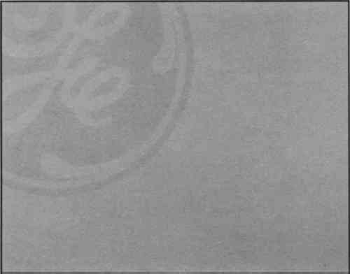
4 NONDESTRUCTIVE IDENTIFICATION TECHNIQUESCHEMICAL WATERMARKS are indistinguishable from traditional watermarks to the unaided eye. Browning (1977) writes briefly of simulated or chemical watermarks and identification techniques. He cites beta radiography and short-wave ultraviolet illumination as methods that can distinguish between traditional and chemical watermarks. Short-wave ultraviolet radiation (180–280 nm) is the easiest method of identification. Chemical watermarks appear dark and dull because components of the watermark absorb the short-wave ultraviolet radiation rather than fluorescing with these wavelengths of light (fig. 5). It is important to inspect both sides of a paper for a chemical watermark, as the marks are better seen on the side of impregnation.
Stationery papers often contain bluing agents or optical brighteners, which fluoresce brilliantly, offering a contrast to the chemical watermark and aiding in the identification process. Chemical watermarks on unbrightened papers lack this contrast and can be harder to see. Fortunately, these marks can be distinguished by using illumination from long-wave ultraviolet sources (300–400nm). Under long-wave ultraviolet illumination, the marks on unbrightened papers appear lighter than their surroundings; they fluoresce as an opaque white in contrast to a dark, nonfluorescing background. Since the traditional watermark is made of the same material as the rest of the paper, it will react like the paper to ultraviolet light, whether or not the paper has been optically brightened. Short-wave ultraviolet radiation is more damaging to the human eye than the long-wave ultraviolet radiation commonly used by conservators. Protective glasses should always be worn when using short-wave ultaviolet radiation, and one should never look directly at the light source. Grenz rays at 7kV were used to radiograph two traditionally and two chemically watermarked papers.2 The traditional watermarks and the paper formation were faithfully recorded on film. Only the paper formation is noticeable on the film exposed under the chemically watermarked papers. This phenomenon occurs because the traditional watermark is a physical manipulation of the paper, whereas the chemical watermark acts more like a size in the paper. Since low-energy X-radiography will not detect organic resins, the chemical watermark is not recorded on film. Rantanen (1989) suggests that an identification can be made under the microscope using raking reflectance illumination. The indented or embossed nature of the traditional watermark will produce shadows. In contrast, variations in surface gloss will sometimes reveal the chemical watermarks as sitting on the surface of the paper. While the differences are easily observed on newer papers, an aged artifact may have been flattened through use, repair, or storage, making positive identification difficult. Placing a blue filter over the light source enhances the effectiveness of this technique. With the filter, the chemical watermarks appear dark on an even surface, while traditional watermarks appear the same color on an uneven surface. The sensitivity of the observer's eye and familiarity with these subtle characteristics is essential when relying on visual microscopic identification techniques. |