A STUDY OF THE DETERIORATION OF EGYPTIAN LIMESTONE SCULPTURES.M. Bradley, & A.P. Middleton
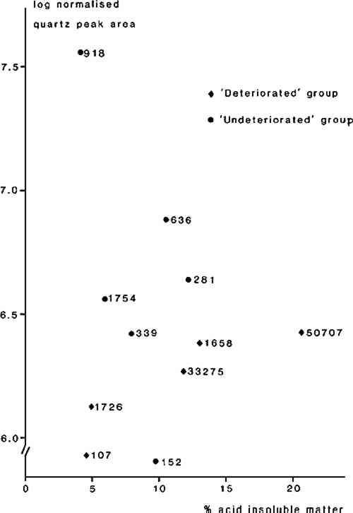
3 RESULTS3.1 Soluble salt analysisTESTS OF THE AQUEOUS extracts for the presence of sulphate ion were negative for all samples. Both chloride and nitrate were present in the “deteriorated” samples from Thebes/Abydos, but in several of the “undeteriorated” sculptures only chloride ions were detected, the level of nitrate ions being below the limit of detection (ca. 1ppm). Statistical analysis using a non-parametric test 17 showed that the nitrate levels tend to be significantly higher in the “deteriorated” group of sculptures but there was no statistical difference in chloride levels between the two groups. The soluble salt levels determined for sculptures from the Cairo area are similar to those for the “undeteriorated” group from Thebes/Abydos. The sculptures from El Bersha were found to have very low levels of both chloride and nitrate. The results are given in Table 2. TABLE 2 Summary of Analytical Results 3.2 Acid insoluble matter determinationThe results of the acid insoluble matter determination are also given in Table 2. The samples from El Bersha have very low acid insoluble fractions whilst those from Cairo and Thebes/Abydos generally have high acid insoluble fractions, indicating differences in the mineralogy of the stones used at the various locations. Although the mean acid insoluble fraction level for the “undeteriorated” group from Thebes/Abydos is lower than for the “deteriorated” group of sculptures there is considerable overlap in the results and the two groups could not be distinguished by statistical tests. 3.3 Identification of acid insoluble fraction (XRD, EDXA)The X-ray diffraction traces obtained on the acid insoluble fractions showed differences between the objects; three broad categories of residue were recognized. These appeared to correlate with the geographical provenance of the sculptures. The results are summarized in Table 2. There was considerable similarity between the acid insoluble fractions obtained from the “undeteriorated” and “deteriorated” sculptures from Thebes/Abydos, and the XRD results indicated that they generally consist of a mixture of non-expanding clay and quartz. Analysis of the clay in the SEM, using EDXA, showed that in most samples it is a magnesium silicate, very low in aluminum, consistent with its identification as either a member of the sepiolite-palygorskite group or as a low aluminum smectite such as hectoraite or stevensite.19,20 The XRD data, particularly the apparently non-expanding nature of the clay, suggest that it is probably an aluminum-poor member of the sepiolote/palygorskite group, since smectite minerals would have been expected to expand on exposure to ethylene glycol vapor. In a few samples, those from EA 33275 and EA 636, the XRD traces indicated that the clay was not of this type, and analysis using EDXA showed that these clays contained aluminum, potassium and iron in addition to magnesium and silicon. Examination of the XRD traces indicated that, in general, the quartz content of the acid insoluble fraction in the “undeteriorated” sculptures from Thebes/Anydos was higher than in residues from the “deteriorates” sculptures. This was tested for the samples from Thebes/Abydos (exception EA 805 for which insufficient sample was available) by determining the quarts peak areas (101 peak) and plotting values normalized for sample weight against the percentage of acid insoluble fraction in the limestone (Figure 6). Statistical analysis of these data, using a non-parametric test, confirmed the significance (at the 95% level) of this difference between the two groups of sculptures.
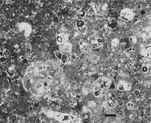
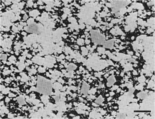
X-ray diffraction analysis was also carried out on stone powder from some of the sculptures from Thebes/Abydos. The traces obtained showed the presence of dolomite in the samples, and normalized peak area measurements (104) peak were obtained for dolomite, as described above for quartz. Although initial inspection of the values suggested that more dolomite was present in the samples from the “deteriorated” group of sculptures, subsequent statistical analysis did not confirm this. X-ray diffraction patterns from the acid insoluble fraction of the sculptures from Saqqara and Giza indicate that they consist mainly of opal-CT, a metastable form of silica,18 sometimes with minor amounts of quartz and/or clay. The very low acid insoluble fractions of the limestone from El Bersha were identified as being mainly quartz. 3.4 Petrographic examinationPetrographic Examination of the polished thin sections indicated that most of the samples are fine grained micritic limestones containing a few fossil fragments (fossilifereous micrites, 21), although some are highly fossiliferous biomicrites (Figure 7). In general it was not possible to observe the non-carbonate fraction in any of the sections in detail, although some EA 33275 from Thebes exhibits form birefringence due to the strongly developed preferred orientation of the clay platelets. The limestones from Thebes and Abydos generally show evidence for partial dolmitisation in the form of small rhombohedra of dolomite (Figure 8). No significant differences were observed between the “deteriorated” and “undeteriorated” groups of sculptures from Thebes/Abydos.
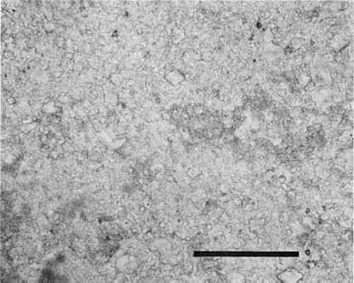
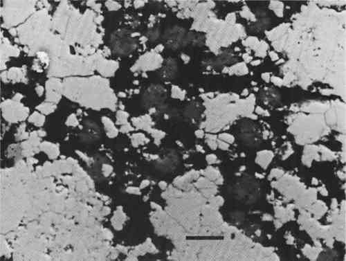
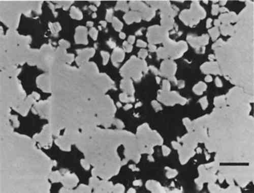
3.5 Scanning electron microscopyThe polished thin sections and fracture surfaces from the sculptures were examined in the SEM. Although in the fracture surfaces detail such as the presence of sodium chloride crystals, grain morphology and distribution and morphology of the non-carbonate fraction were visible, most useful information was derived from the polished thin sections. The observations revealed significant differences between the sculptures, dividing them into three groups which correlate with the geographical provenance. The sculptures from Thebes/Abydos are characterized by the presence of subhedral to euhedral rhombohedra of dolomite in a fine grained micritic matrix of low magnesium calcite (Figure 9). In these samples porosity and the non-carbonate, clay fraction are apparently fairly uniformly distributed throughout the stone. In fracture surfaces the clay could be seen as coatings of flakey or fibrous material on the surface of the calcite grains. The sculptures from Giza and Saqqara have a radically different texture to those from Thebes/Abydos, with islands of relatively solid calcite separated by areas of higher porosity containing lepispheres of silica (opal-CT) (Figure 10). A similar texture was shown by the sculptures from El Bersha (Figure 11), but in these the acid insoluble fraction, present at very low levels, was not readily apparent in the SEM.
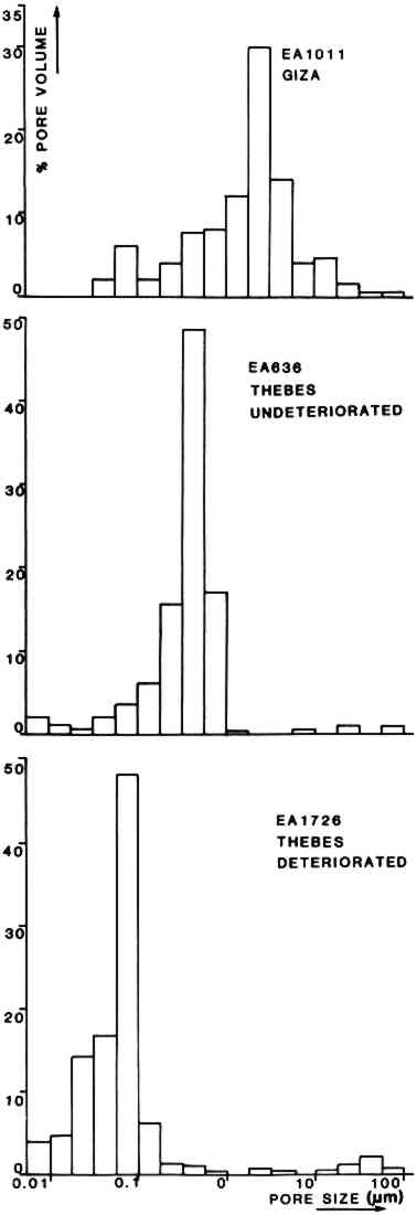
3.6 Mercury PorosimetryHistograms showing pore size distributions of EA 1726 (Thebes, “deteriorated” group), EA 636 (Thebes, “undeteriorated” group) and EA 1011 (Giza) are presented in Figure 12. The data obtained from the determinations are summarized in Table 3. These results suggest that the “undeteriorated” sculptures from Thebes/Abydos have higher total porosity and also higher median pore throat sizes than the “deteriorated” group. Sculptures from the Cairo area have somewhat higher values still of porosity and median throat size. All of the sculptures tested were found to have a high proportion of micropores (i.e. those less than 5 μm in diameter).
TABLE 3 Results of Mecury Porosimetry Determination |