DIGITAL RADIOGRAPHY IN THE ANALYSIS OF PAINTINGS: A NEW AND PROMISING TECHNIQUEA. Everette James, S. Julian Gibbs, Malcolm Sloan, Ronald R. Price, & Jon J. Erickson
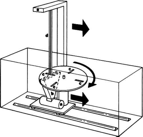
3 Scanned Point Source SystemA third digital radiographic method is the “flying spot” or scanned point source system.6 In this system, the x-ray image is formed by scanning a fine pencil beam of x-rays over the area being imaged (Figure 6). After passing through the object, the
This technique has not been generally available because, until recently, the imaging time was of the order of fifteen seconds, and the spatial resolution was significantly less than with other methods. Although these time and resolution limitations are not so important in radiography of paintings, they are in clinical circumstances. Thus, these machines have not been widely employed in medical facilities. At present, images can be obtained with resolution of about two line pairs per millimeter. In addition, images can be stored and repeated images of the same area can be summed to provide higher quality pictures. Data can be manipulated to change contrast and other characteristics as for the other systems of digital radiography. This technique has not as yet received sufficient use to permit us to evaluate it and to compare it with the other two more widely accepted computer methods. |