DIGITAL RADIOGRAPHY IN THE ANALYSIS OF PAINTINGS: A NEW AND PROMISING TECHNIQUEA. Everette James, S. Julian Gibbs, Malcolm Sloan, Ronald R. Price, & Jon J. Erickson
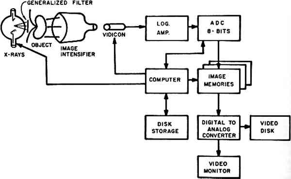
1 Digital FluoroscopyDigital fluoroscopy3, 4 is accomplished using a modified version of the standard radiographic system in which the image is produced by passing x-rays through an object (in this discussion, a painting) to fall on an image intensifier tube acting as the receptor (Fig. 1). In conventional systems, the output of the image intensifier is viewed by a video camera for display or by a film camera system for permanent recording. In
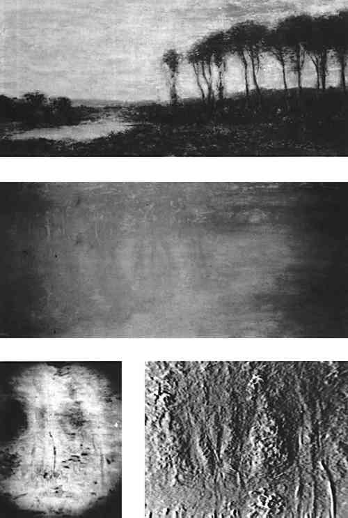
A major advantage of this technique is the instantaneous display of the radiographic image on a television screen. This means that the image may be evaluated for proper exposure and positioning factor while it is being obtained. A second advantage is the very fine spatial resolution that is possible with this imaging device, which is important if one is interested in small details in the images of the paintings. Current image intensifiers have the capability of showing details as small as 0.25 mm in size, allowing the display of fine detail in individual brush strokes in the painting. The major difficulty with the use of this imaging modality is the limited area that can be imaged at one time. The largest image intensifier currently available is only 14 inches in diameter. Thus, the examination of larger areas would require multiple exposures. This system is thus useful for the detailed analysis of a small painting or a portion of a large one, while other digital techniques described subsequently in this communication may be used for overall analysis of works of major size. The digital imaging techniques offers new nondestructive ways to evaluate a painting as well as to compare the components of the work, the artistic technique employed, and the condition of the work. The digital fluroscopic images in Figure 2 show an example of one of the more common techniques of computer image processing, known as subtraction. If two
Other possibilities for this subtraction technique include energy and temporal subtraction. Energy subtraction is based on the well established fact that the attenuation properties of all materials are functions of the energy of the incident x-ray beam. The ability to perform subtraction using digital radiographs obtained at two different x-ray energies may provide additional information in the analysis of paintings. For example, two different pigments may have attenuation characteristics which are nearly identical at a given x-ray energy, making it difficult to differentiate between the two on the basis of contrast in a single image. However, the energy dependence of the attenuation of the two pigments may differ, allowing enhanced contrast by energy subtraction. Temporal subtraction involves the use of two images obtained at different times, with the interval ranging from seconds to years. It may be used to study the deterioration of paintings with time, since only the changes in the attenuation pattern will be displayed in the subtracted image. Further, it could be useful in the authentication of paintings. A digital image of a painting obtained and stored could at some later date be subtracted from a similar image of a suspected copy. Any difference between the two would be exaggerated, making identification of the painting from which the later image was obtained relatively simple and quite precise. |