Analysis and Treatment of a Painting on Vellum with the Aid of a Video Microscopy System
by Julie L. BiggsIntroduction
This paper will discuss the application of a video-microscopy system to the examination and conservation of a painting on vellum. The painting required a course of treatment which was facilitated by the use of the video-microscopy system in the Folger Shakespeare Library's Conservation Department. It is the conservator's aim to address three fundamental issues with respect to the conservation of an artifact: determining the most appropriate method of treatment, acheiving the best results, and documenting the processes thoroughly. Our video-microscopy system was integral to each of these steps, and this project serves as one example of the compatibility between technology and conservation.
Description

Figure 1
The watercolor and gouache painting, acquired by Henry Clay Folger in 1904, measures 19.5 x 14 cm and is solidly adhered to a quarter-cut oak board (Figure 1). The subject of the painting is the Rape of Lucrece (Shakespeare wrote a poem entitled The Rape of Lucrece, which explains why Folger wished to add the painting to his collection of Shakespeariana).1 Lucrece, a Roman matron and image of virtue, is raped by Sextus, the son of Rome's Etruscan ruler, Tarquinius Superbus. Following the rape, Lucrece commits suicide by stabbing herself; her husband and the Brutus family raise a rebellion to avenge her, and the ensuing revolt drives the Tarquins from Rome, ending Etruscan supremacy and leading to the formation of the Roman Republic.
The painting depicts Lucrece in her bedchamber, after she has been raped, on the point of commiting suicide (the painting might be more accurately titled The Death of Lucrece). She has a raised dagger in her hand, and her flighty posture and agitated expression create a sense of drama; there are tears running down her face. In the background, through an open doorway, a group of men can be seen running, hands raised in the air, towards the bedroom. Two dogs are yelping and barking in the fore and middle ground.
This vivid scene is highly stylized and romanticised. There is a sense of anguish for the distraught victim yet there is an absence of horror, for Lucrece, though poised with the dagger, has yet to perpetrate the deed. Perspective is used to great effect: the pattern of the floor tiles leads the viewer's eye through the bedchamber door. The open door and the raised drape in the upper right of the painting allow the audience to assume a kind of presence as onlookers. The detail of the interior - the furniture, coverlet, vessels on the floor and plates on the shelves - conveys a sense of realism and proportion. The dramatic contrast between Lucrece's violation and the orderly domestic interior produces a disarming effect on the viewer. There is a black-painted border around the picture, decorated with gold fronds, vines, and flowers. On the bottom edge of the border is a series of inscriptions that read, "J Bêr," "Mathematic," and "1724."
The Video-Microscopy System
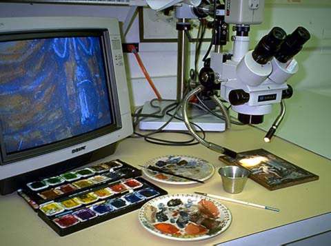
Figure 2
The Folger Conservation Department's video-microscopy system (Figure 2), designed by Micro-Optics, incorporates a Meiji trinocular stereo microscope equipped with a universal boom-stand with tilting mount and a Sony Trinitron high-resolution closed-circuit TV system. 2 The fiber-optic light source is contained in a separate transformer unit that allows the light intensity to be adjusted. The system has a magnification range of x 7 to x 45 through the eyepiece; the monitor magnifies the image approximately x 10 more.3.
Examination and Condition
The painting was framed against glass, and for the past two years has been hanging in a climate-controlled environment, exposed to artificial light from fluorescent tubes. Before 1993 it hung on a wall where it was exposed to sunlight.
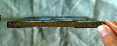
Figure 3
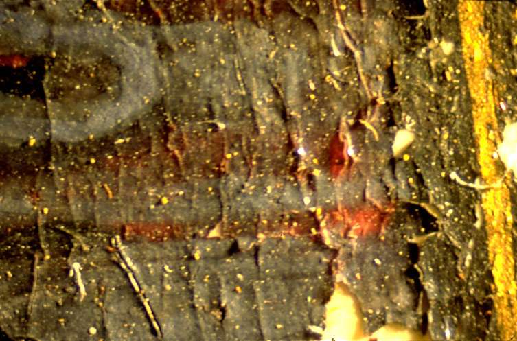
Figure 4
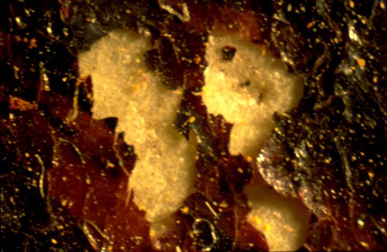
Figure 5
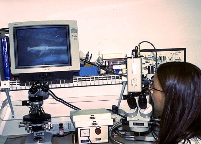
Figure 6
Initial examination showed that the vellum had partially lifted from the oak board (Figure 3). Closer scrutiny revealed cracks and fissures across the entire surface of the painting (Figure 4). In many areas pigment had completely detached from the vellum support, and some parts of the painting were severely damaged (Figure 5).
With the video-microscopy system it was possible to look at these areas in detail over an extended period of time. Observation of the different layers of pigment showed that the paint had lifted more in areas where it was thicker. The density of applied paint and the limited cohesion between the paint binder and the smooth surface of the vellum had resulted in detachment of media. In addition, certain colors seemed to be more vulnerable than others; for example, the gray paint used for the grouting between the floor tiles had flaked off in many parts, while the tiles themselves were virtually undamaged.
It was during this stage of examination that previous restoration work was identified which had not been visible to the naked eye. No restoration has been done since the painting has been in the Folger Collection, so some damage to the piece occured prior to 1904. It was also at this stage that a detail of the original artist's technique was revealed: two contiguous areas of loss expose the vellum support beneath, as well as a red pigment that was commonly used by artists for preparatory sketches. Such sketches were drawn with a diluted ink or pigment. A thin application would have been more readily absorbed by the vellum and was therefore less likely to become detached; a thicker application would tend to sit on the vellum surface. In this case it is clear that the red pigment did not detach, while the overlying paint layer has flaked off.
Pigment samples were extracted with a tungsten needle and mounted to glass slides prepared with Cargille Melt Mount. Slides were viewed under the polarizing microscope at x 40 magnification. The pigments identified were yellow ochre (from the canopy behind the bed), lead white (from the bed linen), madder lake (from the red bedspread), and smalt (from the blue counterpane).
Consolidation
The first phase of the treatment was consolidation of the image. I chose gelatin as the consolidant, which in preliminary tests had proved most effective in securing loose and cupping layers of paint. Furthermore, it was well suited to a precise, localized application and could be readily stored and diluted. The shiny surface that is often seen after consolidation with gelatin results from overapplication. The video-microscopy system was fundamental to controlling the amount of consolidant applied, thus reducing the risk of surface sheen.

Figure 7
We began with a 2 percent gelatin solution that was diluted to achieve optimal wicking. With a sable brush (no. 5/0), the gelatin solution was applied to the lifted pigment via the reticular cracks in the paint surface and along the edge of each loss. The very fine point of the brush enabled accurate placement of the consolidant. On the color monitor it was possible to follow the flow of the gelatin as it wicked underneath the lifted paint layer or fissure (Figure 6). In areas where the paint was thicker and there was greater damage, I made a second application of gelatin.
Compensation of Image
After consolidation, it was necessary to determine how to proceed with inpainting. The video-microscopy system was a useful visual tool in the consultation process between conservators and curators; it was possible to present specific examples of ethical questions on the color monitor and to discuss the treatment options at the same time. The parts of the painting which had been restored earlier were of particular interest: they form a part of the history of the artifact; on the other hand, some of the work was poorly executed and had since been damaged. Because of these considerations, I scrutinized each segment independently.
Inpainting areas of loss proceeded using watercolors, with the addition of methyl cellulose as a binder and thickener. Using the video-microscopy system, I was able to accurately match and apply color to minute areas for several hours at a time. Figure 7 shows the artifact upon successful completion of treatment. Losses were fully compensated and the media secured.
Documentation
All conservation work that alters the existing object in some way must be well-documented. Generally this documentation takes the form of photographs or slides; that is to say, stills of the artifact, before and after treatment has been completed. One unique feature of a video-microscopy system is that it can be used to record video images; multiple computer applications are also possible. Using this method, we were able to produce a video that shows detailed, step-by-step procedures of consolidation and inpainting. This video will serve as documentation and as a teaching tool.4
Conclusion
There are several ways to treat a painting with detached and friable, cracked media. The video-microscopy system is effective for the conservation of a small painting with widespread structural damage and a larger painting with only localized problem areas. Working under magnification can reveal historic details of a painting that are otherwise invisible and enhances manual precision during treatment. The color monitor allows the conservator to work in consultation with others, and to prolong the time spent on treatment yet reduce the risk of eye fatigue and neck strain. Finally, the equipment has the advantage of expanding documentation options that can be done while treatment is proceding.
Acknowledgements
I owe a great debt of gratitude to J. Franklin Mowery, Head of Conservation and Preservation at the Folger Library, who not only inspired me to embark on this project but was extremely generous with his twenty years of knowledge. Mentors are rare these days. My sincere thanks to Philip Weissman of Micro-Optics for his advice on the product, to Steven Kostant for his video expertise, and to Mary Tonkinson for agreeing to edit the manuscript at the drop of a hat.
References
Bykova, G. Z. "Medieval Painting on Parchment: Technique, Preservation and Restoration," Restaurator, 14:3 (1993): 188-97.
Hall, James. Hall's Dictionary of Subjects in Art, rev. ed. (London: John Murray, 1995).
Harley, R. D. Artists' Pigments c. 1600-1835, 2d ed. (London: Butterworth Scientific, 1982).
Harris, William H., and Judith S. Levey, eds. The New Columbia Encyclopedia, (New York: Columbia University Press, 1975).
Inoue, Shinya. Video Microscopy, (New York: Plenum Press, 1989).
Mayer, Ralph. A Dictionary of Art Terms and Techniques, Apollo Edition (New York: Thomas Y. Crowell Co., 1975).
Paper Conservation Catalogue rev. ed. (Washington, D.C.: Library of Congress, 1994).
Roy, Ashok, ed. Artists' Pigments; A Handbook of Their History and Characteristics Vol. 2 (Washington, D.C.: National Gallery of Art, 1993).
Notes
1. Further information on provenance and ownership prior to 1904 was unavailable at the time of publication. E. A. Seemaan lists a "J. Bêr" (or "Jacobber") in Thième-Becker Künstler Lexikon (Leipzig, 1951), and gives 1786 as the year of his birth. The painting is dated 1724, so it cannot be the work of this artist.
2. Micro-Optics Precision Instruments, Philip Weissman, President , P.O. Box 425, Fresh Meadows, N Y 11365.
3. Our system is custom-configured. Adapters can reduce or magnify the image from the microscope to the monitor according to specifications.
4. A five-minute long version of the video was edited for presentation of this paper at the Book and Paper Session of the AIC Conference 1995 so that the audience could see treatment of the painting taking place.
Julie L. Biggs
Senior Paper Conservator
Folger Shakespeare Library
Publication History
Received: Fall 1995
Paper delivered at the Book and Paper specialty group session, AIC 23th Annual Meeting, June 4-1, 1995, St. Paul, Minnesota.
Papers for the specialty group session are selected by committee, based on abstracts and there has been no further peer review. Papers are received by the compiler in the Fall following the meeting and the author is welcome to make revisions, minor or major.