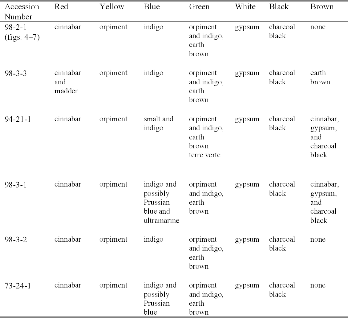
TECHNICAL STUDY OF ETHIOPIAN ICONS, NATIONAL MUSEUM OF AFRICAN ART, SMITHSONIAN INSTITUTIONErica E. James
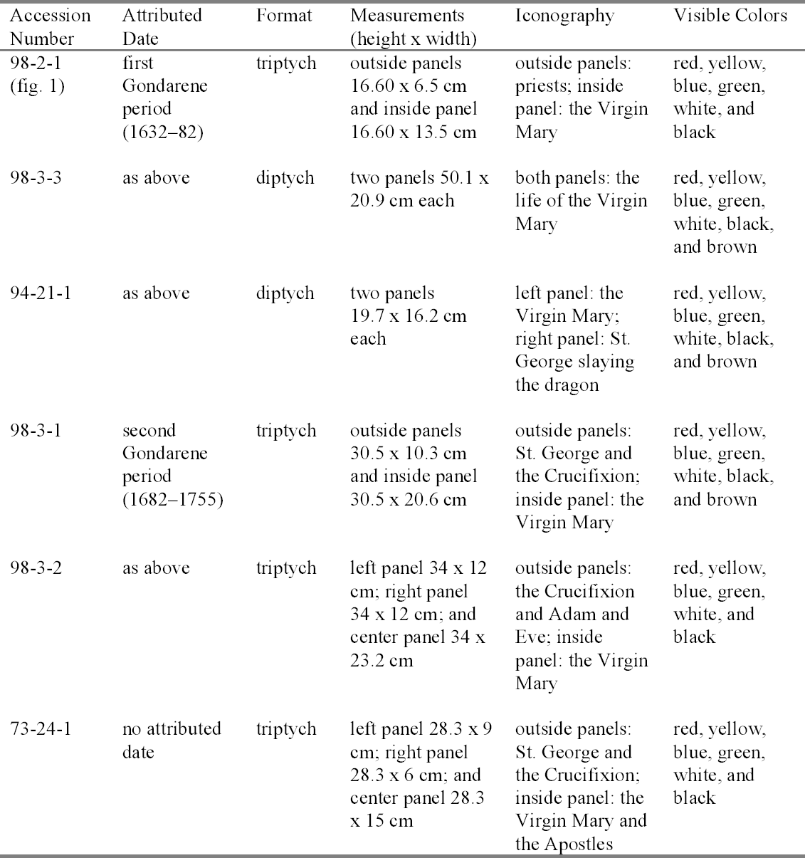
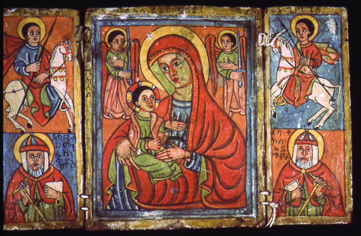
ABSTRACT—This article describes a technical study of six Ethiopian icons at the National Museum of African Art, Smithsonian Institution, and includes a brief synopsis of the history of Ethiopian painting. The preliminary research involved visual and photographic examination using ultraviolet fluorescence and infrared color photography as well as x-ray radiography. Following this initial examination, dispersed pigment samples and cross sections were analyzed using polarized light microscopy. In addition, the cross sections and loose samples were analyzed using scanning electron microscopy with energy dispersive spectroscopy capabilities, x-ray fluorescence, Fourier transform infrared spectroscopy, and x-ray diffraction. The technical study clarified the techniques and materials of Ethiopian icons. The paint layers contained the following pigments: cinnabar, orpiment, indigo, smalt, Prussian blue, terre verte, gypsum, charcoal black, and earth brown. In addition, gypsum was identified as the main component in the ground layer. The binding medium in the pigment and ground was characterized as proteinaceous. This technical study provides insight into icon production in Ethiopia from the 17th to 19th centuries. TITRE—Une �tude technique d'ic�nes �thiopiennes au National Museum of African Art (mus�e national d'art africain), Smithsonian Institution. R�SUM�—Cet article porte sur une �tude technique de six ic�nes �thiopiennes au mus�e national d'art africain, Smithsonian Institution, et comprend un bref r�sum� de l'histoire de la peinture �thiopienne. � l'�tape pr�liminaire de la recherche, on a proc�d� � l'examen visuel et photographique des peintures au moyen de la fluorescence d'ultraviolets, de la photographie infrarouge et de la radiographie. Suivant ce premier examen, on a analys� des �chantillons de pigments en dispersion et des coupes transversales par microscopie en lumi�re polaris�e. De plus, on a analys� les coupes transversales et des fragments par microscopie �lectronique � balayage coupl�e � un spectrom�tre de rayons X, spectrom�trie de fluorescence des rayons X, spectroscopie infrarouge � transform�e de Fourier et diffraction des rayons X. Cette �tude technique nous a permis de mieux comprendre les techniques et les mat�riaux des peintures �thiopiennes. Dans les couches picturales on a identifi� les pigments suivants: cinabre, orpiment, indigo, smalt, bleu de Prusse, terre verte, gypse, noir de charbon et terre brune. De plus, la pr�paration est � base de gypse et le liant des couches picturales et de la pr�paration est de nature prot�ique. Cette �tude technique donne un aper�u de la production des ic�nes en �thiopie du 17i�me au 19i�me si�cle. T�TULO—Estudo t�cnico sobre �cones et�opes, National Museum of African Art, Smithsonian Institute (Museu Nacional de Arte Africana, Instituto Smithsonian). RESUMO—Este artigo descreve um estudo t�cnico sobre seis �cones et�opes pertencentes ao National Museum of African Art, Smithsonian Institute (Museu Nacional de Arte Africana, Instituto Smithsonian), e inclui uma breve sinopse sobre a hist�ria da pintura et�ope. A pesquisa inicial englobou exames visuais e fotogr�ficos utilizando fotografias por fluoresc�ncia no ultravioleta, fotografias a cores no infravermelho bem como raios-X. Em seguida a estes exames iniciais, amostras dispersas e cortes estratigr�ficos de pigmentos foram analisados utilizando microscopia com luz polarizada. Al�m disto, os cortes transversais dos pigmentos e amostras avulsas foram analisados usando microsc�pio de escaneamento por el�tron com capacidade de espectroscopia electr�nica dispersiva, fluoresc�ncia por raios-X, espectroscopia de infravermelhos transformada de Fourier e difrac��o de raios-X. O estudo t�cnico esclareceu as t�cnicas e os materiais utilizados na produ��o dos �cones et�opes. As camadas de pintura continham os seguintes pigmentos: cin�bio, ouro-pimento, �ndigo, esmalte, azul da Pr�ssia, terra verde, gesso, preto de carv�o e castanho terra. Al�m disto, o gesso foi identificado como o principal componente da camada de base. A liga dos pigmentos e da camada de base foi caracterizada como sendo de origem proteica. Este estudo proporciona a compreens�o da produ��o de �cones na Eti�pia desde o s�culo XVII at� ao s�culo XIX. TITULO—Estudio t�cnico de �conos et�opes, National Museum of African Art, Smithsonian Institution (Museo nacional de arte africano, Instituci�n Smithsonian). RESUMEN—Este art�culo 1 INTRODUCTIONThe rich cultural heritage of Ethiopia has been dominated by Christianity, as reflected in Ethiopian icons. The imagery they depict follows traditional Christian themes, and the Virgin Mary is the most popular story and image (Chojnacki 1973). The Virgin Mary is the primary subject in all six of the Ethiopian icons at the National Museum of African Art (NMAfA), Smithsonian Institution (fig. 1). During the 15th century, Ethiopian icons were single panels, diptychs, or triptychs. In the 16th century new construction techniques were introduced by craftsmen who migrated to Ethiopia. These techniques typically involve the use of a wooden secondary support, sometimes a cloth primary support, a ground layer, paint layers, and sometimes a varnish. During this era, at least three European painters are recorded as living in Ethiopia: the Italians Gregorio Bicini and Nicola Brancalion, and the Portuguese Lazaro de Andrade. Their life dates are unknown. The 17th and 18th centuries in Ethiopian painting are described as the Gondarene era. Gondar was the capital of Ethiopia, established on the northern shores of Lake Tana. Here a cultural center gave rise to a style fusing Western models with Ethiopian iconographic ideals. The first Gondarene period occurred from 1632 to 1682; the second Gondarene period occurred from 1682 to 1755. Table 1 lists the Ethiopian icons in the NMAfA collection, including three attributed to the first Gondarene period and two to the second. One icon is not presently attributed to a particular time period. The research and technical data presented here are one of the first attempts to characterize and/or identify the techniques and materials of Ethiopian icons. The first steps in the technical study included examination utilizing ultraviolet light, infrared imaging, and x-ray radiography techniques. Pigment analysis followed, utilizing both polarized light microscopy (PLM) and various instrumental techniques, including scanning electron microscopy with energy dispersive spectroscopy capabilities (SEM-EDS), x-ray fluorescence (XRF), Fourier transform infrared (FTIR) spectroscopy, and x-ray diffraction (XRD). 2 CONSTRUCTION OF ETHIOPIAN ICONSThe Ethiopian icons consist of a wooden secondary support, cloth primary support, a ground layer that adheres the secondary to the primary support, and a paint layer. Six icons in the NMAfA collection are constructed in this manner, although four of the icons lack a cloth primary support. Earlier research indicates that the secondary supports were constructed of wood either from the olive tree or the wanza tree, a type of cedar (Chojnacki 1973). The boards of each icon are usually hewn out of the same tree. The board is taken from the center of the tree, or pith, and the woodwork is done while the wood is still green and pliable. One board is split down the middle to achieve the two panels. Often the panel is chiseled out so that a raised border surrounds the icon on four sides. The raised border design is the earliest type of framing element recorded in European panel paintings, dating to the 13th century (Heydenryk 1963). This raised border design is evident in all the icons at NMAfA. The cotton primary support is adhered to the secondary support in the niche formed by the raised borders. Initially, the wood was primed with a ground layer before the final paint layer was applied, but starting in the 17th century a cloth, usually cotton, Previous field and anecdotal research indicates cinnabar, orpiment, indigo, gypsum, and charcoal pigments were typically used in painting the icons (Weihs 1973). The pigments were bound in a proteinaceous binder, most likely animal glue, producing a distemper medium. Cross sections taken during this current technical study revealed a systematic, deliberate application of paint layers. Finally, earlier research has also indicated that icons created previous to the 17th century were typically unvarnished. Those created later were given a vegetable lacquer coating containing aloe (Weihs 1973). The icons at NMAfA appear to be unvarnished.
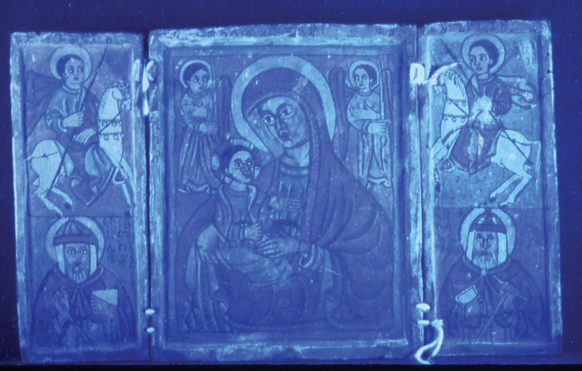
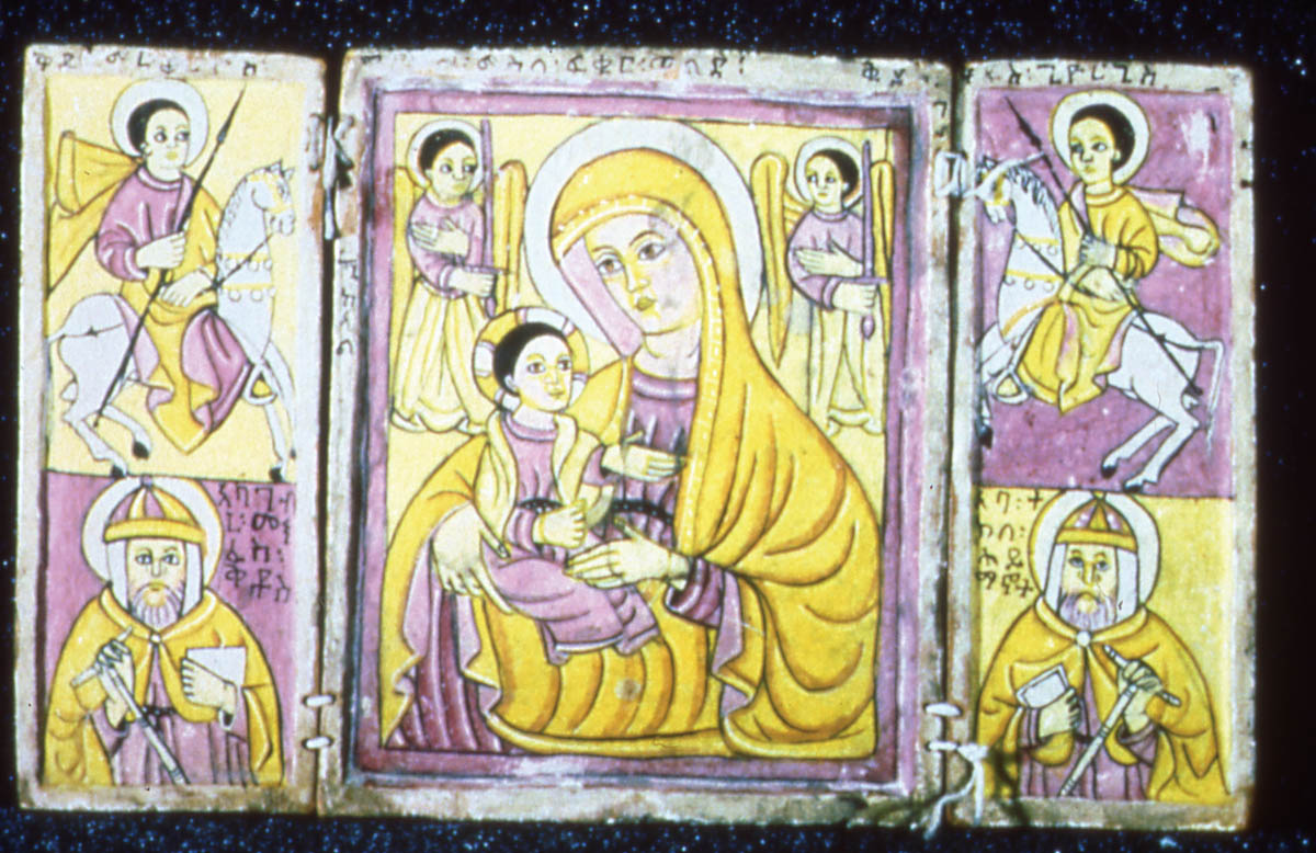
3 TECHNICAL STUDY: VISUAL EXAMINATIONThe first step in documentation was before-treatment photography with examination of the recto surface in normal and raking light and the verso surface in normal light. This photodocumentation was completed using tungsten lighting and Kodak Ektachrome color reversal film ASA 160. In addition, Plus-X Pan black-and-white print film ASA 125 was used to document the recto surface of each icon using normal light. Ultraviolet fluorescence examination was accomplished using tungsten lighting, Kodak Ektachrome color reversal film ASA 160, and a Kodak no. 2B Wratten Gelatin filter. Figure 2 illustrates the fluorescence typical of the icons. The purpose of the UV fluorescence examination was to ascertain the presence of varnish on the surface, to look for the presence of a pink fluorescence often associated with a madder lake paint, and to detect any retouching fluorescence. In visible light, the icons appeared to be unvarnished, and this finding was confirmed with ultraviolet fluorescence examination. One diptych (acc. no. 98-3-3) appeared in visible light to contain madder. Although ultraviolet light examination did not confirm this suspicion, madder was identified on this icon later through dispersed pigment analysis. Retouching was detected on acc. no. 73-24-1 along the lengthwise split in the center panel. Infrared reflectography (fig. 3) and x-ray radiography were also used to detect any losses and/or underdrawings in the paint layer and to discern variable densities of the pigment particles composing the paint layer, respectively. Infrared reflectography (film-based) was accomplished using tungsten lights, Kodak Ektachrome Professional Infrared EIR film (ASA 100/ Process E-6), and a Kodak no. 12 Wratten Gelatin filter. At the NMAfA, IR examination was conducted in Ektachrome slide format. The infrared technique detected only underdrawings that had already been seen during visible examination, thus providing no additional information concerning possible underdrawings not visible to the naked eye. Overall, however, the infrared technique did emphasize how linear and stylized the images in the icons are. Almost all the figures in the icons are outlined with black lines. During x-ray radiography, top views of all the icons were taken with Kodak ReadyPack x-ray film under the following conditions: 70 KV (kilovolts)/5 ma (milliamperes)/116cm TFD (total focal distance
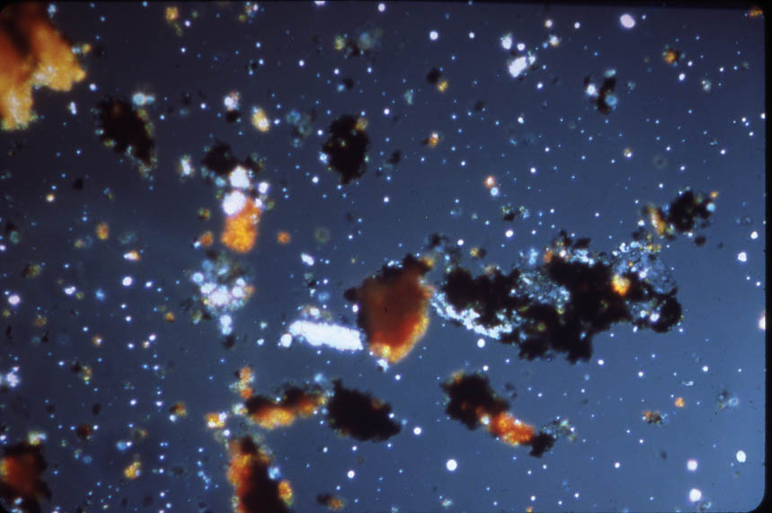
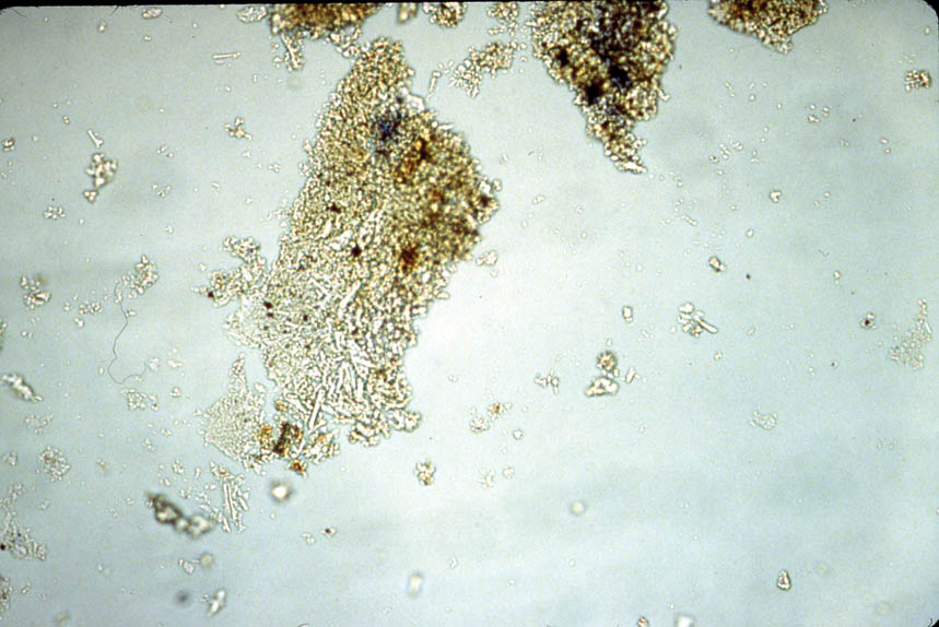
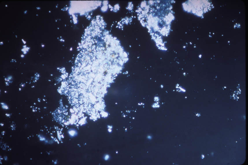
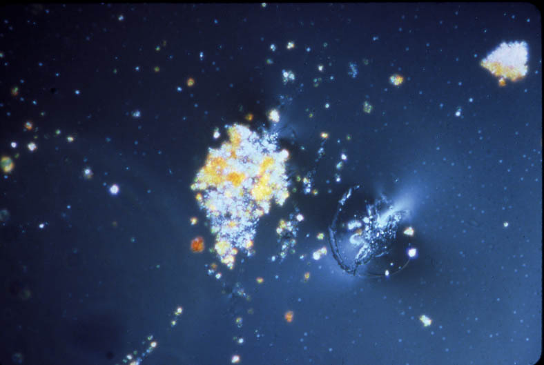
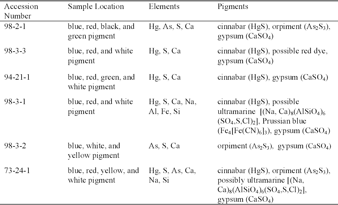
In viewing the x-radiographs of the icons, the goal was to compare the relative densities of the paints on each icon to elucidate materials employed. The areas that were painted red appeared more radio-opaque than other areas due to the density of the red pigment. In acc. nos. 98-2-1, 94-21-1, 98-3-1, 98-3-2, and 73-24-1, the density of one pigment was predominant in the red areas of the paint layer. Cinnabar is mercuric sulfide (HgS) and is the only radio-opaque pigments typically used on the Ethiopian icons. Later analysis by polarized light microscopy did identify the pigment as cinnabar. The x-radiographs revealed no significant deviations in image composition or reconstruction from the final appearance of the icons. 4 TECHNICAL STUDY: EXAMINATION OF PIGMENTSPrior to this technical study, some technical notes on the pigments used for Ethiopian icons had been published (Weihs 1973). To understand each palette, representative colors were sampled on each icon. The individual icons were considered as whole units; thus it was not deemed necessary to sample each panel in a diptych or triptych. 4.1 POLARIZED LIGHT MICROSCOPYThe pigments were examined with dispersed pigment analysis, and each pigment sample was compared to a reference slide. Photomicrographs of dispersed pigments samples are shown in figures 4–7. Pigment samples were removed with a scalpel or needle and mounted with Meltmount mounting medium (refractive index 1.662). The pigment samples were analyzed using the NMAfA Zeiss Axioplan Pol Universal polarized light microscope using transmitted light in bright field and crossed polarization with a magnification range of 100 to 1000x. Polarized light microscopy revealed a consistent palette (table 2): cinnabar (HgS) to create red; orpiment (arsenic trisulfide, As2S3) to create yellow; indigo (C16H10N2O2) to create blue; a mixture of orpiment and indigo to create green; gypsum
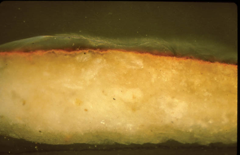
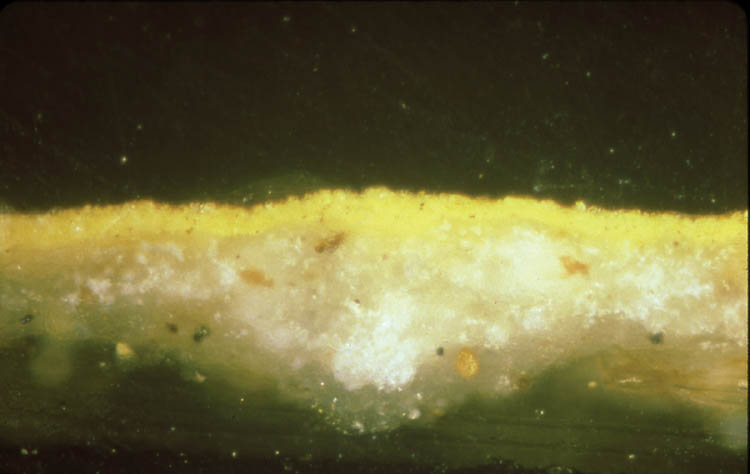
In addition, cross sections mounted in Bio-Plastic polyester resin were examined in order to understand the construction of the paint layer. Figures 8 and 9 are photomicrographs from acc. nos. 98-3-1 and 98-3-2. They both demonstrate the basic direct layering technique used by the icon painter. Figure 8 has a ground layer composed of gypsum and a paint layer composed of cinnabar. Figure 9 has a ground layer composed of gypsum and a paint layer composed of orpiment. 4.2 SCANNING ELECTRON MICROSCOPY WITH EDS CAPABILITIESBoth loose samples and cross sections were then prepared for analysis utilizing scanning electron microscopy with energy dispersive spectroscopy. The SEM instrumentation used in analysis was a JEOL JXA-840A scanning electron microscope with a Tracor Northern (now Noran) TN-5502 energy
In sampling the icons for SEM analysis, cross sections were taken from areas that had the most colors converging in one area. If a color was missed, then a loose fragment was removed for analysis. Despite this consideration, only some of the pigments were able to be isolated for each sample. The SEM-EDS pigment identification results are detailed in table 3. SEM-EDS confirmed the presence of elements that are indicative of cinnabar (mercury and sulfur), orpiment (arsenic and sulfur), and gypsum (calcium and sulfur). The variations to these results included a red sample from acc. no. 98-3-3 with a high organic content, indicating the possibility of a red dye. This result was supported by the PLM identification of madder. Black, green, and blue pigments are difficult to determine by SEM-EDS when their coloration is due to organic compounds. In green areas of the icons, most of the colors described are optical mixtures utilizing orpiment, indigo, and an unspecified earth brown that, depending on the amount added, deepens the green hue. Only in acc. no. 94-21-1 is a pure green pigment, terre verte or green earth, observed and only within a mixture of the other colors. Oral history research indicates that
4.3 X-RAY FLUORESCENCEThe blue areas of the icons were complex in their identification. Although the majority of the blues were indigo, smalt was observed in acc. no. 94-21-1. An attempt was made to confirm the presence of cobalt, the coloring component of smalt, utilizing x-ray fluorescence at the Freer/Sackler Conservation Laboratory. The XRF instrumentation used was the Omega Five x-ray fluorescence instrumentation (a modified Spectrace 6000) with an open-beam architecture. The tube voltage and current were 30 kV and 0.99 mA, respectively. The x-ray beam was directed at the robe of the Virgin Mary. No cobalt was observed in the spectrum of the blue in acc. no. 94-21-1, but the observation of smalt was isolated to one dispersed pigment sample of a pale blue colorant taken from the robe area. Thus, smalt was not the primary 4.4 FOURIER TRANSFORM INFRARED SPECTROSCOPYThe second question in the blue portions of the icons concerned the possible presence of Prussian blue. PLM indicated the possible presence of Prussian blue in the blue paint samples of acc. nos. 98-3-1 and 73-24-1. Since indigo was seen primarily in the other icons, the possible presence of Prussian blue was unexpected. It is difficult, however, to differentiate Prussian blue from indigo using PLM due to their similar optical characteristics, and a precise identification was not possible. Samples from blue areas in all of the icons were analyzed by Fourier transform infrared spectroscopy (FTIR) analysis. The FTIR instrumentation used in analysis was a Mattson 4326 Upgrade Fourier Transform Infrared Spectropho-tometer/SpectraTech IR Plan microscope. The spectra were scanned at 16 or 8 wavenumbers resolution for varying times—3 to 12 minutes. The carbon-nitrogen triple bond (cyano group) of Prussian blue has a distinct absorption at 4.8 microns (2085 wavenumbers). This absorption was observed only in acc. no. 98-3-1. In addition, acc. no. 98-3-1 tested positive for Prussian blue during spot testing1. Prussian blue was not detected in acc. no. 73-24-1 by FTIR analysis or spot testing. The spectra for both of the blue samples indicated two absorption bands in the 1450 to 1500 wavenumber region. Indigoid dyes tend to have two absorption bands in this region. Two such absorption bands are detectable in five of the samples, with the exclusion of acc. no. 98-3-3. The sample for acc. no. 98-3-3 was too difficult to isolate from other materials and impurities in the sample, and so neither Prussian blue nor indigo was detectable. A summary of the FTIR results for the blue pigments is presented in table 4. 4.5 X-RAY DIFFRACTIONThe final question in the blue portions of the painting was the possible detection of ultramarine by PLM in acc. nos. 98-3-1 and 73-24-1 by SEM. This pigment was not observed initially in multiple dispersed pigment samples of blue pigment from
For XRD analysis, each sample was mounted onto the tip of the glass fiber with a rubber cement–toluene mixture. The samples were run on a Philips PW-1820 spectrometer with Ni-filtered Cu K (alpha) radiation at 45 kV and 45 mA for six hours each. Kodak DEF-392 direct exposure film was used to record the diffraction patterns. XRD analysis of these samples showed no evidence of ultramarine. 5 TECHNICAL STUDY: BINDER CHARACTERIZATIONLoose samples were taken from each icon and submitted to the Smithsonian Center for Materials Research and Education for FTIR analysis utilizing solvent extraction. Selected particles were extracted using ethylene dichloride or ammoniacal ethanol, which are effective in isolating the organic material from other inorganic compounds. The primary constituent of the samples, including both paint and ground layers, is proteinaceous. Earlier research indicates that the binder typically used was an animal glue (Weihs 1973). The confirmation of the proteinaceous binder explains the matte quality of the paint layers and corroborates their solubility parameters as detailed in earlier work by the author (James 1999). 6 CONCLUSIONSEthiopian icons have a history that dates back to the 15th century, when the techniques of their construction were introduced by European craftsmen who migrated to Ethiopia (Chojnacki 1973). These techniques typically involve the use of a wooden secondary support, sometimes a cloth primary support, a ground layer, paint layers, and sometimes a varnish. The above technical study characterizes and/or identifies the materials used to construct the icons in the National Museum of African Art's permanent collection. Initial examination utilizing ultraviolet light, infrared imaging, and x-ray radiography techniques revealed that the icons were unvarnished, that no underdrawings were disparate from the final composition, and that the icons contained relatively dense red pigments. Pigment analysis followed, utilizing both polarized light microscopy and various instrumental techniques including scanning electron microscopy with energy dispersive spectroscopy capabilities, x-ray fluorescence, Fourier transform infrared spectroscopy, and x-ray diffraction. These techniques identified cinnabar, orpiment, indigo, smalt, Prussian blue, terre verte, gypsum, charcoal black, and earth brown in the paint layer. In addition, gypsum was identified as the main component in the ground layer, and the binding medium in the pigment and ground was characterized as proteinaceous. In conclusion, the research and technical data presented here are one of the first attempts to characterize the techniques and materials of Ethiopian icons. ACKNOWLEDGEMENTSThe author would like to thank the Kress Foundation for support of this project. A special thanks to Janet Douglas, conservation scientist, and LaHoma Lee, conservation intern, at the Conservation Laboratory, Freer Gallery of Art/Arthur M. Sackler Gallery, Smithsonian Institution, as well as, at the Smithsonian Center for Materials Research and Education, Roland Cunningham, senior painting conservator, Walter Hopwood, analytical chemist, and Camie Thompson, analytical chemist, for analytical assistance with the project. Finally, the author would like to thank Stephen Mellor, chief conservator at the National Museum of African Art, Smithsonian Institution, for his continued support of this project from its inception in June 1998 through its completion in August 2001. NOTES1. The spot test used is found in Berrie 1997. When positive, the test indicates the presence of iron (III), an indicator of Prussian blue. The test enables Prussian blue to be distinguished from ultramarine (natural or synthetic), phthalocyanine blue, and indigo. A stainless steel needle was cleaned with 1:1 ethanol: water, and then the samples were taken. The sample was placed on a slide, and a drop of 4 M sodium hydroxide was added by pipet. A positive result is indicated by a change in color from blue to brown. The blue color can subsequently be restored by the addition of a drop of dilute hydrochoric acid. REFERENCESBerrie, B. H.1997. Prussian blue. In Artists' pigments: A handbook of their history and characteristics, vol. 3, ed. E. W.FitzHugh. Washington, D. C.: National Gallery of Art. 191–217. Chojnacki, S.1973. Ethiopian paintings. In Religious art of Ethiopia,ed. W.Raunig. Stuttgart: Institut fur Auslandsbeziehungen. 33–62. Heydenryk, H.1963. An inquiry into the enhancement of painting.NewYork: James H. Heineman. James, E.1999. Changes in place, changes in context: The conservation of an Ethiopian icon. AIC postprints.American Institute for Conservation 27th Annual Meeting, St. Louis. Washington, D. C.: AIC.69–72. Weihs, F.1973. Some technical details concerning Ethiopian icons. In Religious art of Ethiopia,ed. W.Raunig. Stuttgart: Institut fur Auslandsbeziehungen. 299–305. Wion, A.2001. Personal communication. Mus�e de l'Homme, Paris, France. FURTHER READINGChojnacki, S.2000. Ethiopian icons: Catalogue of the collection of the Institute of Ethiopian Studies, Addis Ababa University. Milan: Skira Editore S. p. A. FitzHugh, E. W.1997. Orpiment and realgar. In Artists' pigments: A handbook of their history and characteristics, vol. 3, ed. E. W.FitzHugh. Washington, D. C.: National Gallery of Art. 47–80. Grissom, C. A.1986. Green earth. In Artists' pigments: A handbook of their history and characteristics, vol. 1, ed. R.Feller. Washington, D. C.: National Gallery of Art. 141–67. Hartman Samet, W.1994. The philosophy of aesthetic reintegration: Paintings and painted furniture. In Painted wood: History and conservation,ed. V.Dorge et al. Los Angeles: Getty Conservation Institute. 412–24. Langmuir, E., et al. 1978. Ethiopia: The Christian art of an African nation. Salem, Mass.: Peabody Museum of Salem. Muhlethaler, B., and J.Thissen. 1993. Smalt. In Artists' pigments: A handbook of their history and characteristics, vol. 2, ed. A.Roy. Washington, D. C.: National Gallery of Art. 113–30. Piechocinski, M. N.1989. The iconography of Ethiopia: A review of the styles, themes, techniques, and influences in an historical perspective. Sacred Art Journal10(3): 95–112. Plesters, J.1993. Ultramarine blue: Natural and artificial. In Artists' pigments: A handbook of their history and characteristics, vol. 2, ed. A.Roy. Washington, D. C.: National Gallery of Art. 37–65. Portell, J. D.1986. African red pigments. The care and preservation of ethnological materials: Symposium 1986, ed. R.Barclay. Ottawa: Canadian Conservation Institute. 119–29. Scholz, P. O.1992. Orbis Aethiopicus. Albstadt, Germany: Karl Schuler Publishing. Wolde, S.1986. The preservation and conservation of art works in present-day Ethiopia. Proceedings of the First International Conference on Ethiopian Art. London: Royal Asiatic Society. 131–35. SOURCES OF MATERIALSBio-PlasticWard's Natural Science P.O. Box 92912 Rochester, N.Y. 14692-9012 (716) 359-2502 AUTHOR INFORMATIONERICA E. JAMES graduated from the Winterthur/University of Delaware Program in Art Conservation in 1999. Following graduation, she completed a Getty Fellowship at the Baltimore Museum of Art, Baltimore, Maryland, and a Kress Fellowship at the National Museum of African Art, Washington, D.C. She is currently in private practice in painting conservation in Lancaster, New Hampshire. Address: P.O. Box 349, Lancaster, N.H. 03584
 Section Index Section Index |