SHORT COMMUNICATION: MICRO-RAMAN IDENTIFICATION OF BLOOM FORMED ON A HISTORICAL PRINT ARTIFACTVINCENT OTIENO-ALEGO, JENNIFER HODGEMAN, & DUDLEY C. CREAGH
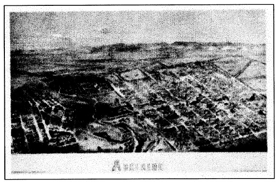
1 INTRODUCTIONThe original black-and-white bird's-eye print forms part of a large pictorial collection of the Adelaide City Archives, Adelaide, South Australia. Originally drawn by A. C. Cooke (1836-1906) (Kerr 1992) and engraved by L. Y. S. Calvert (1828-1913) in 1876 (Kerr 1992), the bird's-eye print, or Calvert Panorama, as it is often referred to, depicts an aerial view of Adelaide city with its picturesque Mount Lofty ranges in the background (fig. 1). This particular print appeared as a supplement in the Adelaide Illustrated News of July 1876 and has been used extensively as a resource for heritage and historical inquiries. During a recent general inspection of the print, some whitish powder was observed on its surface as well as on the overlying glazing. The print also exhibited overall mottling and local areas of brownness. While the print was being deframed, a powdery material was observed to be loosely attached to the print surface, and it could easily be dislodged by gentle brushing. An annotation referring to a previous conservation treatment in 1972 was also noted. Unfortunately no details were given of the treatment. Therefore, before commencement of the new treatment, a scientific analysis of the loose solids was undertaken to assist in identifying previous treatment. In particular, this type of analysis assisted in resolving health and safety issues related to the potential toxicity of the powder (which might have contained pesticides). Analysis of bloom on art objects has used a number of techniques, including differential scanning calorimetry (DSC) (Williams 1989; Burnstock et al. 1993; Singer et al. 1995), infrared microscopy (IR) (Williams 1989; Burnstock et al. 1993), gas chromatography coupled to mass spectrometry (GC-MS) (Burnstock et al. 1993; Singer et al. 1995), scanning electron microscopy (SEM) (Burnstock et al. 1993), and energy-dispersive X-ray analysis (EDX) (Burnstock et al. 1993; Singer et al. 1995). In this study, Raman microscopy was used to analyze white powder trails observed on the bird's-eye print. This technique has been widely used in the past few years to identify pigments present in art objects (e.g., Bruni et al. 1999; Clark 1999; Derbyshire and Withnall 1999; Edwards et al. 1999; Coupry 2000; Otieno-Alego 2000). Similar to the more frequently used technique of IR, the Raman technique also involves the irradiation of a sample by a monochromatic light source (usually a laser) to produce a particular combination of the Raman bands that reflects the vibrational modes typical of the material under examination. However, because of differing quantum mechanical selection rules, IR and Raman spectra are not identical but are said to be complementary (Schrader 1995).That is, vibrations that are strong in a Raman spectrum are usually weak in an IR spectrum and vice versa. Qualitatively, antisymmetric vibrational modes and vibrations due to polar bonds (such as O-H, N-H, C=O) generally exhibit prominent IR bands, while Raman tends to emphasize vibrations involving more symmetrical bonds (such as C=C, C-C, S-S) (Schrader 1995).
To the analyst, Raman microspectroscopy offers many benefits in comparison to IR microspectroscopy. The most important are the ease of sample preparation, sampling through glass and plastic, ability to analyze aqueous samples, and the compatibility with fiber optics allowing for remote sampling. Furthermore, the laser beam used in Raman technique can be focused to achieve a high spatial resolution (typically 1 �m), which is 20 times better than the focus achievable with IR radiation (Best et al. 1992). This high spatial resolution facilitates the
|