DIGITAL VIDEO MICROSCOPY: A PRACTICAL VISUAL ANALYSIS TECHNIQUE FOR THE CONSERVATORTED STANLEY
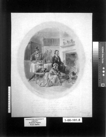
3 3. VIDEO MICROSCOPY APPLICATIONS FOR ROUTINE CONSERVATIONA conservator can take advantage of a number of practical and valuable everyday uses for video microscopy in his or her laboratory. The application of digital video microscopy is not necessarily limited to a few conservation specialty groups but can be used whenever side-by-side visual comparison studies are needed. The following are several practical instances in which I have found digital video microscopy to be an important and valuable tool. 3.1 3.1 SPOT TESTING SOLUBLE PIGMENTSSpot testing pigments is a routine examination activity that is performed by a paper conservator. Spot testing is conducted to determine a pigment's likely reaction to various reagents or liquids before an actual treatment is carried out. Once testing begins, it is always difficult to remember how a very small area, a few millimeters in diameter, truly appeared before testing. Therefore, trying to determine exactly how the pigment is being affected by the application of a reagent is sometimes problematic. The conservator has a powerful tool for this type of situation by using the side-by-side visual comparison capabilities of a digital video microscopy system. For example, a pen and ink wash by Felix Octavius Carr Darley (1822–1888) was brought to my attention by the Graphic Arts Collection curator (fig. 2). Darley worked as an illustrator in Philadelphia and New York City during the 1840s and 1850s (Groce and Wallace 1957). The drawing suffered from a nasty mat burn that obscured the drawing. An aqueous treatment was contemplated for the drawing to reduce the mat burn. The ink wash and paper support had to be spot tested before beginning any treatment to minimize the risk of ink loss and also to predict what sort of aqueous solution would be effective in reducing the staining.
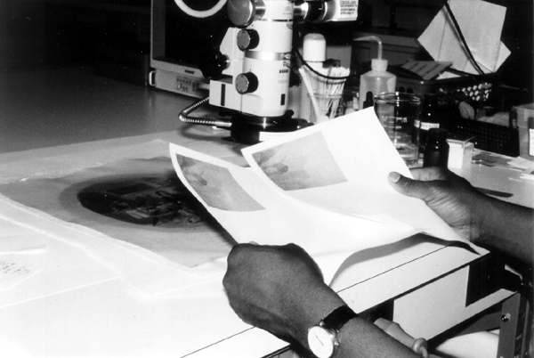
The initial test was to determine a safe and effective aqueous solution for washing the object. A very tiny and obscure area of the drawing was selected for testing. A still image of the area was captured and saved on the computer before testing began. I then brought up a real-time frame image of the area to be tested beside the still-frame image on the computer monitor. Both images were 320 � 240 recorded resolutions. As I tested the area with various solutions of water and absolute ethanol, I could observe the effect of the solutions and compare it to the image of the same untested area on the computer monitor. I could also periodically print out images of the area, tested and untested, as the examination progressed to make visual comparisons of the testing (fig. 3). The images were made part of the treatment documentation in hard copy and computerized form. The same type of testing continued for finding an aqueous solution for stain reduction. Exhaustive testing found that (1:4) absolute ethanol:distilled water solutions were suitable for washing. Further, the same proportions of absolute ethanol: 1% and 2% buffered hydrogen peroxide (for immersion and local applications, respectively) proved to be safe and effective for stain reduction using the same comparison method as before.
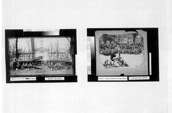
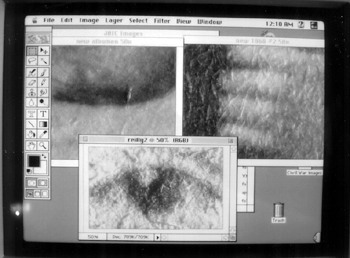
3.2 3.2 IDENTIFICATION OF PHOTOGRAPHSDifferent types of photographic processes have developed since William Henry Fox Talbot's invention in 1839. Some of these processes can be identified through visual examination of the photograph's surface characteristics. A trained eye can pick up the subtle distinctions between one process and another, but differentiating closely related photographic processes is made easier by the use of side-by-side comparison techniques. I received several 19th- and early-20th-century photographic prints for conservation from the Princeton University Archives (Seeley G. Mudd Manuscript Library). Two photographs dated 1868 (Princeton class of 1868) and 1890 (Princeton class of 1890) were of particular interest when their surface characteristics were examined under a stereo microscope (fig. 4). At first I thought the 1868 print may have been a salt print, but, on closer inspection, I found an extremely light coating of albumen. The 1890 photograph also appeared to be an albumen print, but with a much thicker coat. The fibers of the paper appeared distinctly through the albumen layer, and there was no apparent baryta layer, which is indicative of a gelatin print (Reilly 1986). I generated magnified images of the respective surfaces on my computer and displayed them side-by-side (fig. 5). It appeared that the 1868 photograph was most likely an albumen photograph that perhaps had not been commercially prepared. The print had a very distinct textured surface because of an extremely thin coating of albumen over the paper fibers. Both prints were further examined and conserved following their identification.1
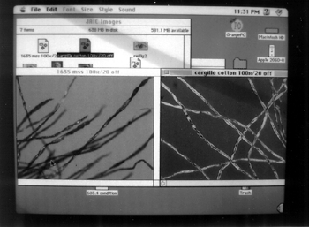
3.3 3.3 FIBER AND PIGMENT ANALYSISDigital video microscopy is a valuable aid in the examination of paper and other types of cellulosic fibers. The morphology of a fiber can be carefully observed using a polarized light microscope. Comparative observations between known and unknown specimens make the task of identification much easier to perform. A fiber sample is placed between a glass slide and cover, and a refractive index indicator medium is added (McCrone et al. 1995). Through polarized light microscopy, specific morphological characteristics of the unknown specimen can be compared to known samples that may lead to the specimen's identification. Examination of the unknown specimen proceeds by analyzing it for such characteristics as size, signs of elongation, extinction, vessel walls, refractive index, etc. There are several very useful sources of reference materials covering the morphological features of fibers, such as C�t�'s Papermaking Fibers: A Photomicrographic Atlas (1980). Using digital video microscopy, a still image of the unknown specimen is obtained and saved in the computer. The unknown specimen on the microscope stage is replaced with a known specimen that approximates or matches the morphological characteristics of the specimen. The known specimen can be compared in a real-time frame on the monitor side-by-side against the saved image of the unknown specimen. Through systematic comparative study, the unknown specimen can be identified (fig. 6). Fiber samples for carrying out comparative analysis studies can be obtained from several sources, such as the Institute of Paper Science and Technology and McCrone Accessories and Components, which also sells Cargille pigment sample kits.
Comparative pigment analysis studies can also be made in conjunction with performing standard examination protocols. Unknown-pigment specimens are compared side-by-side to known samples. Various reference sources for pigment characteristics may be consulted to help in the identification process. Walter McCrone (1982) has published an excellent reference, which has analytical data on a great number of historic artists' pigments. Another excellent source for analytical pigment data is the Artists' Pigments series (Feller 1986; Roy 1993; Fitzhugh 1997) published by the National Gallery of Art, which covers a wide range of historic pigments. |