DIFFERENCES IN IMAGE TONALITY PRODUCED BY DIFFERENT TONING PROTOCOLS FOR MATTE COLLODION PHOTOGRAPHSSYLVIE PENICHON
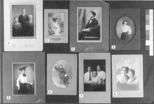
10 ENERGY DISPERSIVE X-RAY SPECTROMETRY (EDS) ELEMENTAL ANALYSISThe versatility of the matte collodion process and the infinite variety of tones it provided were repeatedly praised in the literature of the period. The second part of this study consisted of the elemental analysis of several matte collodion photographs in order to establish a relationship between toner and final color of the print. Energy dispersive x-ray spectrometry (EDS) is a nondestructive analytical technique for determining the elemental composition of a material. The technique itself is non-destructive, but it is often thought of as destructive because the analysis is usually done on samples taken from objects rather than on the object itself. Other nondestructive methods, such as x-ray fluorescence (XRF), where samples are not placed in vacuum during the operation and prints of virtually any size can be analyzed, are usually preferred by conservators for elemental analysis. There are two main advantages of EDS microanalysis over XRF for this project. First, EDS is usually done at a lower voltage The EDS detector measures the energies of the characteristic x-rays produced by each element in the material. These x-rays are produced after bombardment of the sample with a beam of electrons, usually from a scanning electron microscope (SEM). By measuring the energies of the different x-rays produced, EDS can qualitatively identify which elements are present in a material. By counting the number of each energy x-ray produced, EDS can also provide a quantitative analysis of the composition. Eight matte collodion photographs (fig. 8) were selected from study collections so the widest variety of image tonalities would be represented. Because matte collodion prints are sometimes difficult to distinguish from matte gelatin prints through visual examination only, a spot test was applied on every photograph to confirm that the binder was collodion. The test consists of applying a drop of deionized water on the photograph and letting it sit for a minute before it is dried with a blotter. The wet area is then observed under a microscope. Unlike gelatin that swells when it comes in contact with water, collodion does not absorb water and will not show any change. Sampling and analyses were performed by Mark Wypyski, associate research scientist at the Sherman Fairchild Center for Objects Conservation of the Metropolitan Museum of Art, New York. The photographs were sampled in the darkest areas of the image, where more image components (silver and toning metal) are expected to be found. Samples measuring approximately 0.25 square mm were taken using a steel scalpel and mounted onto carbon sample mounts with conductive carbon paint. The analyses were done using a Kevex model Delta IV EDS attached to
TABLE 3. ELEMENTAL ANALYSIS OF MATTE COLLODION PHOTOGRAPHS All the samples showed large amounts of sulfur and barium, ranging from 55 to 77% of the sample's total composition. This finding indicates that a great amount of baryta (barium sulfate and gelatin) layer was taken together with the binder during the sampling of the photograph. Aluminum and silicon were present in variable amounts in all the samples. These elements constitute “background noise” to any material being analyzed; their presence can be discounted. In addition to the silver image, three of the images analyzed revealed the presence of gold only; four showed a combination of gold and platinum; one had platinum only. Print 1, showing three adults, is a very slightly gold-toned photograph. Gold represents only 2% of the total composition of the sample and 8% of the image material. The tone of the print is brownish red and resembles the color of an untoned POP. This color is not often seen in matte collodion prints. The sample taken from Print 2, showing a standing woman with a hat, contains a greater percentage of gold than silver. The photograph displays a reddish purple tone. Print 3, showing a seated man, was gold toned to a dark purplish color. The large amount of gold in the sample, 65% of the image material, indicates a thorough substitution of the silver image. Print 4, showing a girl with a ribbon in her hair, is a gold-platinum-toned photograph. There is much more gold than platinum in the sample and the photograph displays black neutral tone. Silver remains the main component of the image material indicating short gold and platinum baths. The same observation can be made for Print 5, a gold-platinum-toned photograph of Print 6, showing a seated girl, is a gold-platinum-toned photograph. This time, gold and platinum are in equal amounts in the sample. The color of the print is a neutral, somewhat washed gray. Print 7, showing three children, is a gold-platinum-toned photograph that displays a neutral black hue. The amount of platinum in the sample is twice the amount of gold. Print 8, showing two young women, was toned with platinum only and displays a brownish gray tone. |