LASER STAIN REMOVAL OF FUNGUS-INDUCED STAINS FROM PAPER
HANNA MARIA SZCZEPANOWSKA, & WILLIAM R. MOOMAW
3 RESULTS OF LASER TREATMENT OF FUNGAL STAINS
At the appropriate laser power and pulse frequency, YAG laser treatment at 532 nm was very effective in removing the stains associated with two of the four fungi studied, Alternaria solani(figs. 3a–b) and Penicillium notatum(fig. 3c). In both of these cases, the stains were completely removed after 3 to 10 laser pulses. Inspection under both the light microscope (fig. 3) and at higher power using the scanning electron microscope (fig. 4b) revealed no damage to the paper fibers in the treated area. The stains associated with Chaetomium globosum and Fusarium oxysporum appeared to be unaffected by the laser treatment.
Fig. 3a.
Cultivated black stain of Alternaria solani. The white area indicated by the arrow is the region cleaned by the laser.
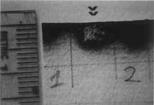 |
Fig. 3b.
Close-up of laser-cleaned area of peper stainde by Alternaria solani, surrounded by remaining stain
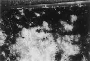 |
Fig. 3c.
Paper stained by Penicillium notatum. Area indicated by arrows has been cleaned by laser irradiation.
 |
What was truly remarkable was the discovery that in the case of Penicillium notatum, the fungal bodies embedded in the paper matrix were removed along with the stain.Figure 4a clearly reveals the fungal spores in a scanning electron micrograph, and figure 4b shows the intact paper fibers and absence of fungal bodies after treatment. The lower power levels of the dye laser were generally found to be less effective in removing stains, but successful dye laser removal of Fusarium fungal bodies was achieved at these power levels after 3 minutes of pulsing at 20 pulses per second (fig. 6b). Additional optimization studies are needed.
Chaetomium globusum is a highly cellulolytic fungus that utilizes cellulose fibers as a carbon nutrient source, turning pliable paper into a pulpy form with a porous, blotterlike structure of low tear strength. With sufficient time and favorable conditions, all cellulose fibers are digested.Figure 5 illustrates Chaetomium globusum–damaged paper. Because of the fundamental damage to the fibers by this fungus, any type of mechanical treatment on the affected area will only cause further rearrangement and deterioration of the paper fibers. Unfortunately, laser treatment under the conditions we used did not appear to be effective in removing Chaetomium stains.
Fig. 5.
(right) Paper fiber structure damaged by Chaetomium globosum Scanning electron micrograph. 40x
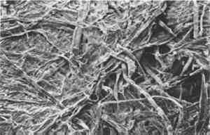 |
In the case of Fusarium oxysporum, laser treatment did not appear to remove stains successfully, but the scanning electron microscope revealed that small holes approximately 5–10 μm in diameter were produced (fig. 6). The holes appear to be where the mycelium or fungal bodies penetrated the structure of the paper and are the voids left behind when the mycelium is removed by the laser light rather than produced by the laser itself. Otherwise, one would expect to see similar holes in fungus-free samples as well. Therefore, in this case the laser treatment removed the colored fungal bodies but not the stain.
Fig. 6a.
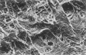
Fusarium oxysporum fungal filaments on paper. Scanning electron micrograph. 124x
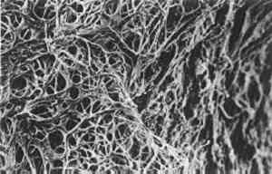 |
Fig. 6b.
Paper fibers following attempted cleaning with dye laser irradiation of Fusarium oxysporum. Although stain was not removed, fungal filaments are gone and small 5–10 μm holes can be seen where they penetrated the paper. Scanning electron micrograph. 124x
 |
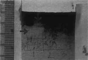
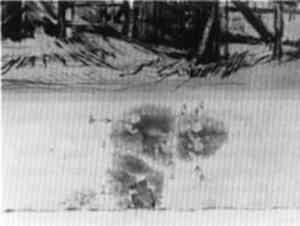
The technique was tested on a print (fig. 1) that had been stained most likely by fungus Alternaria solani. The print was a 19th-century Flemish etching printed on medium-weight wove paper. It was possible to remove considerable amounts of the stain with only 1 to 6 laser pulses without degrading the quality of the paper itself (fig. 7).
Fig. 7.
Laser stain removal on the border of the etching in figure 1. The small light spot was produced by a single laser pulse from the YAG laser, while the clearer, larger spot required six pulses.
 |
To determine possible damage to ink and other pigments, the YAG laser was aimed at an area of heavy black printer's ink on the Flemish print. Thirty pulses at the highest average laser power (1.6 watts) were required to remove a noticeable amount of ink (fig. 8). This number of high-energy pulses is 6 to 10 times higher than that needed to remove fungal stains. Similar power levels were also capable of removing pastel pigment from another piece of paper, but loss of pigment was insignificant when power levels were lowered by 75%.
|