SOME IMPROVEMENTS IN THE STUDY OF CROSS SECTIONSJIA-SUN TSANG, & ROLAND H. CUNNINGHAM
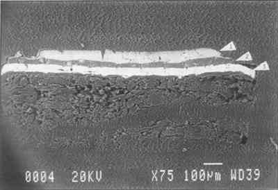
4 APPLICATIONS OF THE METHODTHE ABOVE represents the general scheme of chemical and physical examination and chemical tests that can be applied to thick and microtomed cross sections made from paint samples. Some applications of this scheme and the results we achieved are examined below. Observations made on the cross section also are discussed, together with the analytical results. With the microtome, multiple cross sections can be generated from one paint sample, making it possible to use one sample for many tests. 4.1 STANDARD SECTIONSA series of known pigments and binding media was applied to Masonite and aged for six months in the Conservation Analytical Laboratory's painting conservation studio using the environmental control present in the building (70 � 2�F and 50 � 2% RH). By layering paints of known pigments and binding media on a Masonite board, layered cross sections were created. The test panels were used as controls to test the sensitivity and limitations of the analytical methods described above. These test samples were especially useful in testing and perfecting the sectioning system. Samples mounted in a large cylinder mold were used for thick sectioning, and samples mounted in BEEM molds were used for microtoming. Sample A consists of: 1) Masonite sized with rabbit skin glue; 2) a ground layer of calcium carbonate and rabbit skin glue; 3) a first layer of Rowney tempera cadmium red in a protein and oil medium; and 4) an upper layer of lead white in an egg yolk medium. Sample E consists of: 1) Masonite sized with rabbit skin glue; 2) a ground layer of lead white and rabbit skin glue; 3) a first layer of Rowney tempera ochre color in a protein and oil medium; and 4) an upper layer of zinc white with egg yolk medium. A 15-micron microtomed cross section of sample E was prepared following the procedure described previously. This cross section was then examined using scanning electron microscopy and the EDS system. Elemental analyses confirmed the presence of the pigments. Despite the fact that the blade used for this particular cutting was not very sharp, the clear SEM image presented in figure 2 indicates that the unpolished microtomed surface is suitable for scanning electron microscopy.
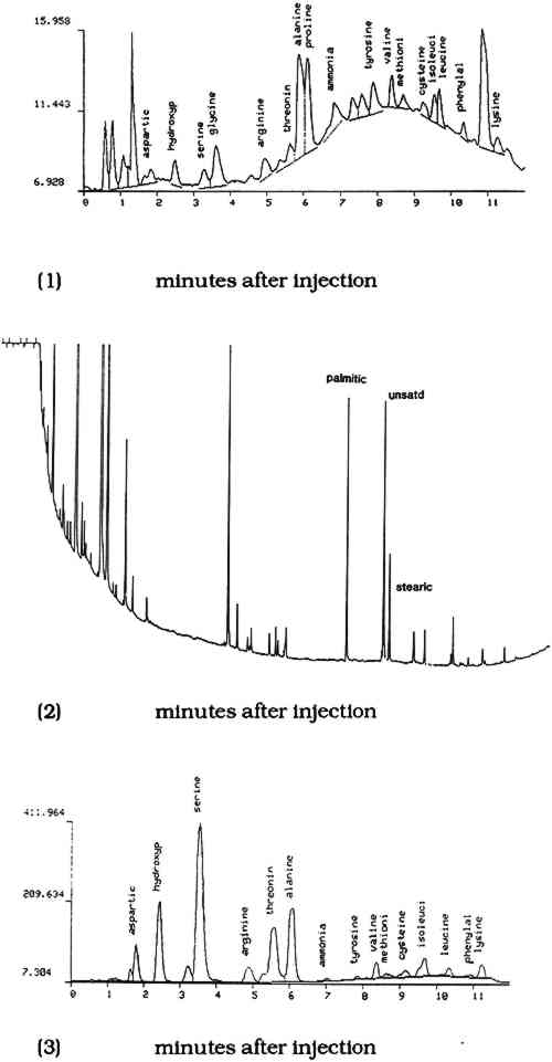
Another microtomed section from sample E was subjected to a fluorescence-staining test for protein, using 0.25% fluorescein isothiocyanate in ethanol. A third microtomed section then was subjected to the fluorescence staining test for oil using solution of 0.25% Rhodamine B in ethanol as the staining reagent. The staining results confirm the presence of the media applied aa described above. Under the stereomicroscope, a small portion of each layer was excavated and used as a sample for GLC identification of oils1 and the HPLC analysis of proteins.2 The ground and secondlayers were tested for protein by HPLC, and the middle layer was tested for drying oils by GLC. Figure 3 represents the chromatograms of each layer. Despite the small size of the sample retrieved from the thin-section, the methodology and instrumentation are sensitive enough for identification purposes.
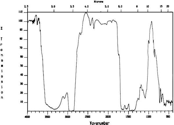
The microtomed section from sample A was subjected to FTIR analysis by transmitted light through the microscope. The microtomed sections were thin enough to allow light to be transmitted through them. The transmission mode was chosen over reflection because existing spectra in the conservation literature are primarily transmission spectra and a large computerized data bank exists for transmission spectra; the reflection mode is still under investigation for its application in conservation (Baker et al. 1988). The Conservation Analytical Laboratory instrument consists of the Spectra-Tech IR-Plan microscope attached to a Mattson Cygnus 100 FTIR. The microscope can be operated in a visible light mode for viewing the sample, with the FTIR beam condensed to focus in on a very small spot. Spectra were collected for each paint layer in the cross section, and they confirm the presence of the material included in the preparation. These results are presented in figure 4. The FTIR spectra of the embedding
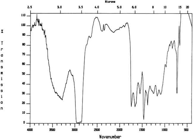
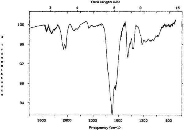
4.2 TECHNICAL EXAMINATION AND STUDY OF AMERICA RECEIVING NINE MUSES, A 1903 PAINTING BY THOMAS DEWING4.2.1 General Description of the Painting and its ConditionThomas Dewing's America Receiving Nine Muses (1903) depicts 10 women dressed in turn-of-the-century ball gowns. America is shown in profile seated in a curule chair at the right of the painting. There are three standing figures to the left and six dancing figures on the right. Overall, light pastel colors such as pink, mauve, and gray are set in a greenish background. This work is of Dewing's last period (1900–1915). It is painted on the piano lid of the 100,000th Steinway piano, that was presented to the White House in 1905, during President Theodore Roosevelt's term of office. The piano case was very ornately gilded, designed by J. D. and J. H. Hunt. The case surface is a painted scrolled foliate border surrounding and connecting shields that represent the 13 original states. A more complete description of the painted surface and its conservation is presented elsewhere (Tsang et al. 1990). The support for the painting is in good condition with no apparent warping, delamination, or other instability associated with the wood of which it is composed. The paint layers also are in very good condition, and it appears that the painting has never been cleaned. There are abrasions in the painting resulting from improper handling of the piano lid. The natural resin varnish layer is discolored and has marred the tonal and spatial quality of the painting. Removal and repair of the abrasions were the objective of the conservation work. 4.2.2 Analysis of the PaintingSolvent tests were conducted using cotton swabs with mild solvents commonly used for removal of natural varnish. The results of these tests indicated a slight potential paint solubility problem since the cotton swabs had a brown coloration. It was very difficult to tell from the swabs whether any of the greens had been affected. Tests of the paint layers and their binding media were then conducted to assess the effect of varnish removal on the paint underneath. First, the cotton swab from the acetone cleaning tests was reextracted with acetone. Any material that dissolved was then cast on a potassium bromide pellet, and an FTIR spectrum was taken. This spectrum indicated the presence of a dammar-like natural resin (fig. 5). There was no indication of other material being present.
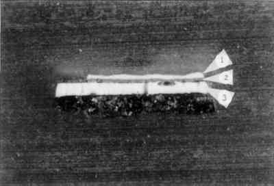
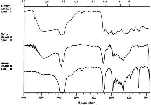
Second, microtomed sections of paint sample were prepared and used for hot-stage melting points, fluorescence-staining, FTIR, and SEM analysis. Examination under the light microscope of the cross section of a sample from the green background indicated that the paint film is composed of a green over a white over a gold layer, with a white gesso layer at the bottom. The top green layer reacted positively for oil using the Rhodamine B fluorescence test. The hot-stage melting point tests indicate that at between 80�C and 90�C a resin like material flows out of the top green layer and the green layer appears more broken up. At approximately 180�C the green color disappears, and at 230�C the white gesso layer turns brown. These color changes are unrelated to the presence of an oil and a wax or resin. The results indicate that the top green layer contains a wax or soft resin and that oil was used as the pigment binding medium. FTIR analysis of a microtomed section also indicates the possible presence of beeswax and oil in the green layer (fig 6). However, this evidence is not conclusive.
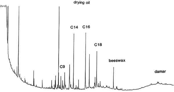
GLC of a portion of this section indicates the presence of oil and beeswax in the green paint layer and the presence of dammar from the varnish on top of the green paint. These results are presented as figure 7.
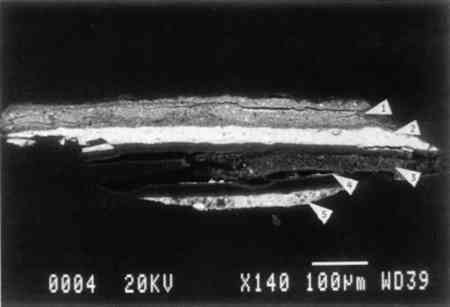
It must be noted that FTIR spectra of the extract from the cotton cleaning swab indicated only the presence of the major component—a dammarlike natural resin. The small amount of beeswax present in the green paint was not detected. FTIR is used in some conservation laboratories as a routine screening test for the safety of the solvent cleaning The hot-stage melting point study of the microtomed cross section also indicates the presence of low-melting components such as a resin-wax in addition to the oil medium of the green paint. This study is simple and useful and can be used as an adjunct to more sophisticated techniques. Fluorescence staining with the oil stain Rhodamine B indicates that drying oil is present in the top green layer. However, our subsequent findings indicate that oil staining alone also cannot be used as the sole source of analysis for a complex mixed media. GLC and GC-MS of the dry paint sample and cotton swabs were used to confirm the presence of beeswax and drying oil. In this case study, the presence of beeswax was not detected by FTIR and fluorescence staining. However, it was detected by the hot-stage melting-point test and by chromatography. A microtomed section from the green background was mounted on a graphite stub using a water-based adhesive prior to carbon coating in preparation for the SEM study. Solvent-based mounting adhesives that might affect the binding medium were avoided. A SEM photograph of the section shown in figure 8. Elemental analysis of this section by EDS shows chromium and zinc in the top layer, indicating the presence of chrome green and zinc white: lead in the second layer indicates lead white, and similarly the third layer contains gold and the fourth iron, indicating iron oxide. Lead white was present in the ground layer.
|