SOME IMPROVEMENTS IN THE STUDY OF CROSS SECTIONSJIA-SUN TSANG, & ROLAND H. CUNNINGHAM
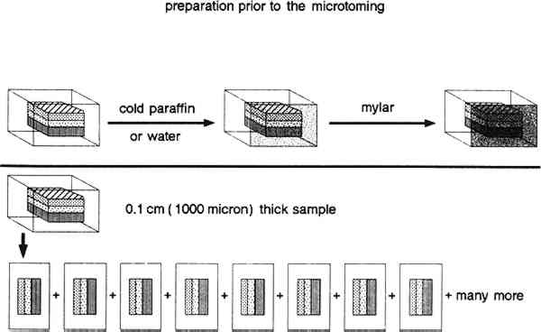
ABSTRACT—This paper outlines techniques developed at the Conservation Analytical Laboratory, Smithsonian Institution, for the preparation of thick cross sections (50 microns and up) and microtomed cross sections (10 microns and up) of small samples from works of art. The sectioning methods discussed here enable us to maximize the information that can be retrieved from samples of paint films. One advantage of the 10-micron microtomed sections is that they are thin enough to be analyzed non-destructively using transmission Fourier transform infrared spectrometry (FTIR). These results can be used as the first material classification step in our analytical materials identification scheme. We can then proceed using destructive chemical and physical tests of thin-sections with greater confidence. The identification of the binding media and pigments from several cross sections is also described. 1 INTRODUCTIONTHE STUDY of cross sections can provide a large amount of precise information about the composition of a very small area from a work of art. It complements other techniques such as x-radiography and examination under ultraviolet and infrared light, which give rather more general information over a large area. Pioneering work on painting cross sections was conducted by Raehlmann, who published his results in 1910. Another important earlier paper was published by Plesters (1956) on cross sections and the chemical analysis of paint samples. It is still widely referred to today. Although the technique has become widespread in conservation laboratories in museums and in conservation training schools, the basic approach of microscopic examination and chemical staining and testing has remained relatively unchanged for many years. During this time, however, techniques of instrumental analysis have changed tremendously. While instrumental analyses have been used to solve other problems in conservation, they have not always been suited to the study of the individual layers in cross sections either because of the limited sample size, or the fact that most instrumental analyses are destructive, or that sample preparation methods were inadequate for the proposed techniques. Consequently, there is a lack of correlation between the technical information gathered from the microscopic study of cross sections and that from instrumental analyses. The use of a microscope for identification of the pigments and media in a cross section is a technique that is accessible and much used by most conservators in most conservation laboratories. Unfortunately, techniques such as gas-liquid chromatography (GLC) and high-performance liquid chromatography (HPLC) are accessible to only a few conservators and conservation scientists in a few conservation laboratories. The paper published by Erhardt et al. (1988) suggested a systematic approach to the instrumental analysis of natural finishes and binding media. That paper is useful as a guide and reference for those who do have access to an analytical laboratory. In that analytical scheme, the suggested first step is to obtain an infrared spectrum of the sample. The use of these spectra makes it possible to narrow the range of materials in a sample by excluding various potential components and to classify in a general way those major components that are present. We describe below our method for providing cross sections thin enough for a transmitted light Fourier transform infrared (FTIR) microscope to obtain infrared spectra of the individual layers in a cross section. This is the first step in linking the study of cross sections directly to other instrumental analyses. In addition, by thinly slicing cross sections, we increase the number of samples that can be used both for microscopic and other analytical 2 PREPARATION OF SAMPLES FOR CROSS SECTIONINGTWO TYPES of samples are prepared, thick sections and microtomed sections. Thick section preparation is used to produce samples from 50 microns and up in thickness. Microtomed section preparation produces samples 10 microns and up in thickness. The type of embedding mold used depends on the size of the sample, type of sample material, and possible identification methods. For thick sectioning, the large round cylinder molds 32 mm diameter � 33 mm high (total volume 20 ml) or small cylinder molds 26 mm diameter � 13 mm high (total volume 5 ml) are used. For the microtome procedure, samples are placed within BEEM molds, which are flat polyethylene-embedding molds, 7 mm wide � 15 mm long � 3 mm thick, tip 2 mm (total volume 0.5 ml) or 9 mm wide � 19 mm long � 6 mm thick, tip 2 mm (total volume 1.0 ml). The embedding material consists of unsaturated polyester resin in styrene monomer with methyl ethyl ketone peroxide as hardener (Buehler Castolite embedding resin). The curing time at room temperature for this system is 6–8 hours, with increasing viscosity noted during in the first 12–14 minutes. A half block is first cast, and the sample is carefully placed in the cylinder or BEEM mold. This is done under a stereomicroscope so the sample can be arranged to provide the best angle for successful sectioning. A tiny dab of cyanoacrylate adhesive can be used at the edge of sample block to secure the sample in its preferred orientation before pouring in the rest of the polyester resin. Cold curing without an elaborate air-evacuating system at room temperature is preferred. A 6 in long blood coagulation tube whose tip has been drawn out to a fine point over an alcohol lamp is used to manipulate the sample and to remove bubbles in the embedding material. 2.1 PREPARATION OF THICK SECTIONS 50 MICRONS AND GREATERThick sections are prepared when 1) pigment particles are larger than 10 micron; 2) a microtome is not available; or 3) larger samples are available. To prepare these sections, a Buehler Isomet low-speed saw is used for sectioning, and a Buehler Minimet grinder is used for polishing. Buehler and other companies also have additional cutting and grinding instruments that can be adapted to this purpose. First, a sample that has been embedded in polyester resin and hardened overnight is slipped out of its disc mold and placed onto a suitable chuck from the cutting machine. The chuck is used to hold the sample firmly in position to prevent both inaccurate cuts and damage to the blade due to movement while sectioning. Various chucks have been designed to meet most sample shape requirements. We use a wafering blade, which consists of a thin metal disc (5 in diameter � 0.015 in thick) having a rim of fine grit diamond grain embedded in a powdered metal matrix. A blade rotation speed of 200–250 rpm is chosen for most of our applications. The desired section thickness is achieved by using a micrometer-controlled cross feed. Samples of an exact thickness can be cut by considering the micrometer setting, the desired section thickness, and a sample loss factor based on blade thickness. Whenever possible, we use only water as the cutting and polishing fluid. We avoid the use of other lubricants because of the difficulty they can cause during subsequent chemical analysis. They can introduce additional matrix components that might confuse the study of original material, especially by FTIR analyses. However, in cases in which water cannot be used, especially when water soluble materials such as glue and gum exist in the cross sections, kerosene can be used as lubricant. A first cut is made to expose the sample, which is embedded near the center of the polyester disc. The exposed sample is then polished before the sectioning proceeds. In addition, the sample is always polished before each new section is taken. In this way, one side of any thin-section slice is always polished; a polished, smooth surface is crucial 2.2 PREPARATION OF MICROTOMED SECTIONS OF 10 MICRONS AND GREATERLeitz Model 1300 and model 1400 sledge microtomes and a Reichert-Jung model 2040 rotary microtome have been used effectively to section paint samples. The specimen is first embedded in the polyester as before, in this case using a BEEM mold, and is trimmed on the Isomet saw to expose the sample. The sample is then placed in the microtome and held by clamps that allow orientation of it in any direction. Section thickness can be set by moving the object into the stationary blade or by having the cutting knife move into the object. Usually, the mechanism is built into the microtome body and moves the object. The advance mechanism is coupled with the cutting action so that each object advance takes place after a cutting action and can be set to advance manually or automatically. The section thickness setting is adjustable to from 1–40 microns. Working with samples from paintings from different periods and of different styles, we found a reasonable and workable thickness for the cross sections to be 10–15 microns. With some older paintings with coarse, hand-ground pigments, it might not be possible to make sections this thin. This thickness was established from working with different matrices of varying hardness and thickness and taking into account the sensitivity and limitations of the microtome and FTIR microscopy. For instance, for an FTIR microscope study, the maximum section thickness that would permit an interpretable transmitted light spectrum was found to be about 20 microns. After the embedded sample is positioned in the sample holder of the microtome, a small piece of Mylar about the size of the tip of the mold is attached to the exposed sample with water or cold paraffin. Because of the complex layering and inconsistent hardness in the layered structure of a painting, we use Mylar or other polyester films to absorb the stress to the sample resulting from cutting it. Our previous unsuccessful attempts to prepare microtome cross sections were related primarily to the unregulated cutting shock which was powerful enough to break the fragile and thin paint layer. A very small amount of cold paraffin is used to ensure the adhesion of the Mylar film. Paraffin is chosen because of its minimal interference in an IR spectrum. At the beginning of the project, we found that excess cold paraffin could cause a problem both in FTIR analysis and a hot stage melting point study. As a result of practice and experience, we have been able to minimize the amount of paraffin we use. Consequently, we applied cold paraffin mostly on the perimeter of the sample, then gently rubbed the Mylar with a smooth metal rod on the sample surface to ensure the use of the minimum amount of paraffin while still promoting adhesion of Mylar to the surface. The sample with the Mylar attached is then cut with the microtome. The sample is kept in contact with the Mylar until the microtomed section is ready to be used. Separating the Mylar from the sample is achieved by dipping it briefly into liquid nitrogen. The resulting thermal shock embrittles the paraffin which aids its removal and helps separate the Mylar from the sample without introducing forces that might tear the microtomed section. In addition, the liquid nitrogen does not introduce impurities that might cause confusion during chemical analysis. Typically, there is no need to polish
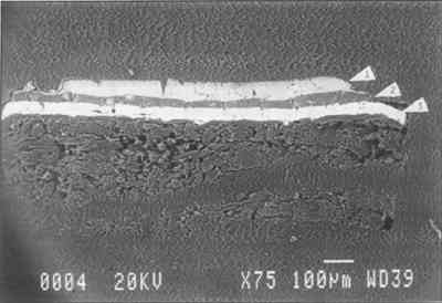
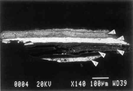
3 METHODS THAT CAN BE APPLIED TO THE STUDY OF THICK AND MICROTOMED CROSS SECTIONSSOME SIMPLE, rather accessible microscopic examination methods (3.1, 3.2, 3.3, 3.4) can be very valuable in identifying and understanding the material; suitable microscopes are available to most conservation laboratories. In many of these tests, however, the cross section is destroyed in the process of analysis. With other methods some portion of the prepared section is used up in the process of testing (GLC, HPLC, XRD and SEM). With FTIR the section is not used up. 3.1 EXAMINATION WITH A STEREOMICROSCOPEThis method permits visual study and photographic recording of the cross section and its layered structure together with observation of its color, texture, thickness, and the pigment particle size. It can provide preliminary identification of the pigments. The solubility studies can be conducted by applying various solvents to the cross section. Characterization (not identification) of the media and overpaint can facilitate decision making for treatment purposes. Staining tests can be conducted for the study of binding media (Johnson and Packard 1971). 3.2 HOT-STAGE MICROSCOPYMicro-melting-point studies can be used to divide the medium into subgroups such as resin, oils, and protein. Thin sections allow faster heat transfer and make the studies more effective. Traditionally, melting points are carried out using much thicker castings. 3.3 FLUORESCENCE MICROSCOPYThis method can be used to identify pigments and conduct staining tests of binding media (Wolbers 1988). 3.4 POLARIZED LIGHT MICROSCOPYWith this method, pigments can be retrieved from the cross section for examination under microscope. 3.5 GAS-LIQUID CHROMATOGRAPHY (GLC) AND GAS CHROMATOGRAPHY–MASS SPECTROGRAPHY (GC-MS)Fragments can be saved from the original sample or samples can be retrieved under a stereomicroscope with needles from the thin section for more accurate identification of binding media. 3.6 HIGH-PERFORMANCE LIQUID CHROMATOGRAPHY (HPLC)Fragments can be saved from the original sample or samples from the thin section can be retrieved for more accurate identification of the amino acids in a protein-binding medium. 3.7 X-RAY DIFFRACTION (XRD)This method provides accurate identification of inorganic pigments. Samples can be retrieved from the cross section. The sample can be reused for another type of analysis if separated from the mounting media. 3.8 SCANNING ELECTRON MICROSCOPY (SEM), WITH ENERGY-DISPERSIVE X-RAY SPECTROMETRY (EDS)The morphology of each layer from the cross section can be studied at high magnification using the SEM, and elements composing each pigment can be identified in each layer of paint using EDS. The SEM sample can sometimes be reused for another type of analysis if it is large enough for repolishing, which is necessary to remove the carbon or gold coating. 3.9 FOURIER TRANSFORM INFRARED SPECTROSCOPY (FTIR)Analysis of an individual layer in the microtomed cross section can be conducted using transmitted light. This analysis provides a determination of the general chemical class of the layer without destroying the cross section and without involving complicated derivatization procedures. FTIR can be used to determine which further analytical techniques would be most appropriate. 4 APPLICATIONS OF THE METHODTHE ABOVE represents the general scheme of chemical and physical examination and chemical tests that can be applied to thick and microtomed cross sections made from paint samples. Some applications of this scheme and the results we achieved are examined below. Observations made on the cross section also are discussed, together with the analytical results. With the microtome, multiple cross sections can be generated from one paint sample, making it possible to use one sample for many tests. 4.1 STANDARD SECTIONSA series of known pigments and binding media was applied to Masonite and aged for six months in the Conservation Analytical Laboratory's painting conservation studio using the environmental control present in the building (70 � 2�F and 50 � 2% RH). By layering paints of known pigments and binding media on a Masonite board, layered cross sections were created. The test panels were used as controls to test the sensitivity and limitations of the analytical methods described above. These test samples were especially useful in testing and perfecting the sectioning system. Samples mounted in a large cylinder mold were used for thick sectioning, and samples mounted in BEEM molds were used for microtoming. Sample A consists of: 1) Masonite sized with rabbit skin glue; 2) a ground layer of calcium carbonate and rabbit skin glue; 3) a first layer of Rowney tempera cadmium red in a protein and oil medium; and 4) an upper layer of lead white in an egg yolk medium. Sample E consists of: 1) Masonite sized with rabbit skin glue; 2) a ground layer of lead white and rabbit skin glue; 3) a first layer of Rowney tempera ochre color in a protein and oil medium; and 4) an upper layer of zinc white with egg yolk medium. A 15-micron microtomed cross section of sample E was prepared following the procedure described previously. This cross section was then examined using scanning electron microscopy and the EDS system. Elemental analyses confirmed the presence of the pigments. Despite the fact that the blade used for this particular cutting was not very sharp, the clear SEM image presented in figure 2 indicates that the unpolished microtomed surface is suitable for scanning electron microscopy.
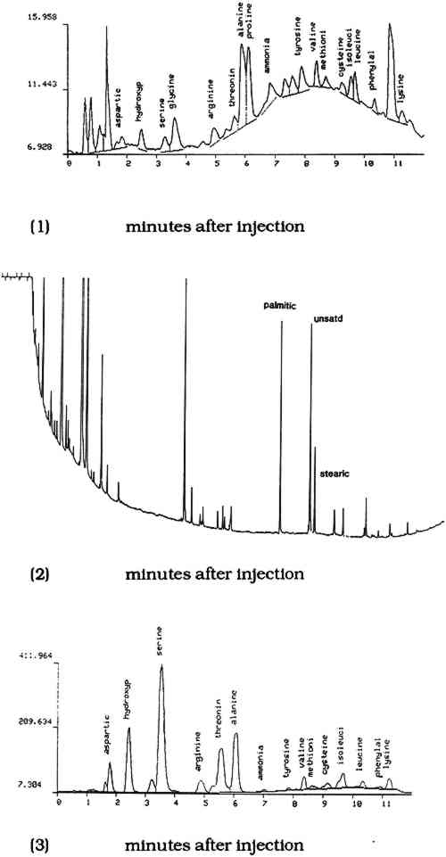
Another microtomed section from sample E was subjected to a fluorescence-staining test for protein, using 0.25% fluorescein isothiocyanate in ethanol. A third microtomed section then was subjected to the fluorescence staining test for oil using solution of 0.25% Rhodamine B in ethanol as the staining reagent. The staining results confirm the presence of the media applied aa described above. Under the stereomicroscope, a small portion of each layer was excavated and used as a sample for GLC identification of oils1 and the HPLC analysis of proteins.2 The ground and secondlayers were tested for protein by HPLC, and the middle layer was tested for drying oils by GLC. Figure 3 represents the chromatograms of each layer. Despite the small size of the sample retrieved from the thin-section, the methodology and instrumentation are sensitive enough for identification purposes.
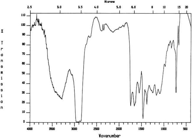
The microtomed section from sample A was subjected to FTIR analysis by transmitted light through the microscope. The microtomed sections were thin enough to allow light to be transmitted through them. The transmission mode was chosen over reflection because existing spectra in the conservation literature are primarily transmission spectra and a large computerized data bank exists for transmission spectra; the reflection mode is still under investigation for its application in conservation (Baker et al. 1988). The Conservation Analytical Laboratory instrument consists of the Spectra-Tech IR-Plan microscope attached to a Mattson Cygnus 100 FTIR. The microscope can be operated in a visible light mode for viewing the sample, with the FTIR beam condensed to focus in on a very small spot. Spectra were collected for each paint layer in the cross section, and they confirm the presence of the material included in the preparation. These results are presented in figure 4. The FTIR spectra of the embedding
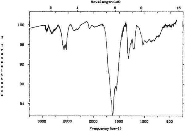
4.2 TECHNICAL EXAMINATION AND STUDY OF AMERICA RECEIVING NINE MUSES, A 1903 PAINTING BY THOMAS DEWING4.2.1 General Description of the Painting and its ConditionThomas Dewing's America Receiving Nine Muses (1903) depicts 10 women dressed in turn-of-the-century ball gowns. America is shown in profile seated in a curule chair at the right of the painting. There are three standing figures to the left and six dancing figures on the right. Overall, light pastel colors such as pink, mauve, and gray are set in a greenish background. This work is of Dewing's last period (1900–1915). It is painted on the piano lid of the 100,000th Steinway piano, that was presented to the White House in 1905, during President Theodore Roosevelt's term of office. The piano case was very ornately gilded, designed by J. D. and J. H. Hunt. The case surface is a painted scrolled foliate border surrounding and connecting shields that represent the 13 original states. A more complete description of the painted surface and its conservation is presented elsewhere (Tsang et al. 1990). The support for the painting is in good condition with no apparent warping, delamination, or other instability associated with the wood of which it is composed. The paint layers also are in very good condition, and it appears that the painting has never been cleaned. There are abrasions in the painting resulting from improper handling of the piano lid. The natural resin varnish layer is discolored and has marred the tonal and spatial quality of the painting. Removal and repair of the abrasions were the objective of the conservation work. 4.2.2 Analysis of the PaintingSolvent tests were conducted using cotton swabs with mild solvents commonly used for removal of natural varnish. The results of these tests indicated a slight potential paint solubility problem since the cotton swabs had a brown coloration. It was very difficult to tell from the swabs whether any of the greens had been affected. Tests of the paint layers and their binding media were then conducted to assess the effect of varnish removal on the paint underneath. First, the cotton swab from the acetone cleaning tests was reextracted with acetone. Any material that dissolved was then cast on a potassium bromide pellet, and an FTIR spectrum was taken. This spectrum indicated the presence of a dammar-like natural resin (fig. 5). There was no indication of other material being present.
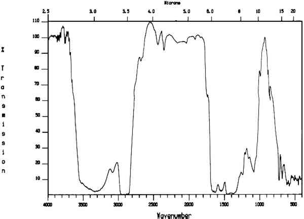
Second, microtomed sections of paint sample were prepared and used for hot-stage melting points, fluorescence-staining, FTIR, and SEM analysis. Examination under the light microscope of the cross section of a sample from the green background indicated that the paint film is composed of a green over a white over a gold layer, with a white gesso layer at the bottom. The top green layer reacted positively for oil using the Rhodamine B fluorescence test. The hot-stage melting point tests indicate that at between 80�C and 90�C a resin like material flows out of the top green layer and the green layer appears more broken up. At approximately 180�C the green color disappears, and at 230�C the white gesso layer turns brown. These color changes are unrelated to the presence of an oil and a wax or resin. The results indicate that the top green layer contains a wax or soft resin and that oil was used as the pigment binding medium. FTIR analysis of a microtomed section also indicates the possible presence of beeswax and oil in the green layer (fig 6). However, this evidence is not conclusive.
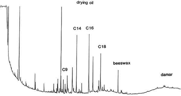
GLC of a portion of this section indicates the presence of oil and beeswax in the green paint layer and the presence of dammar from the varnish on top of the green paint. These results are presented as figure 7.
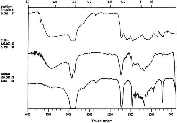
It must be noted that FTIR spectra of the extract from the cotton cleaning swab indicated only the presence of the major component—a dammarlike natural resin. The small amount of beeswax present in the green paint was not detected. FTIR is used in some conservation laboratories as a routine screening test for the safety of the solvent cleaning The hot-stage melting point study of the microtomed cross section also indicates the presence of low-melting components such as a resin-wax in addition to the oil medium of the green paint. This study is simple and useful and can be used as an adjunct to more sophisticated techniques. Fluorescence staining with the oil stain Rhodamine B indicates that drying oil is present in the top green layer. However, our subsequent findings indicate that oil staining alone also cannot be used as the sole source of analysis for a complex mixed media. GLC and GC-MS of the dry paint sample and cotton swabs were used to confirm the presence of beeswax and drying oil. In this case study, the presence of beeswax was not detected by FTIR and fluorescence staining. However, it was detected by the hot-stage melting-point test and by chromatography. A microtomed section from the green background was mounted on a graphite stub using a water-based adhesive prior to carbon coating in preparation for the SEM study. Solvent-based mounting adhesives that might affect the binding medium were avoided. A SEM photograph of the section shown in figure 8. Elemental analysis of this section by EDS shows chromium and zinc in the top layer, indicating the presence of chrome green and zinc white: lead in the second layer indicates lead white, and similarly the third layer contains gold and the fourth iron, indicating iron oxide. Lead white was present in the ground layer.
5 CONSERVATION TREATMENT OF THOMAS DEWING, AMERICA RECEIVING NINE MUSESTHE EXTENSIVE tests we conducted confirm the presence of a small amount of beeswax in the green layer. GLC analysis indicates that some of this wax was removed by cleaning tests of the varnish layer with cotton swabs. Cleaning tests with mild solvents also demonstrated a degree of blanching. This information was sufficient to warrant a decision not to remove the varnish. The varnish had been applied by brush, and uneven drip spots are noticeable on the green background. It was decided to reduce these uneven spots by applying solvent with a laboratory cleaning 6 DISCUSSIONIN THE field of conservation we frequently encounter results that are inconclusive due to the sample size and inability to repeat tests because of the destructive nature of the test itself. By using thick-sectioning and microtome-sectioning technique, we increase the number of available tests samples that can be provided from one small original sample. As demonstrated above, there is no single test that can offer the necessary satisfactory results. To confirm one analysis, a number of different tests must be conducted. Even when sophisticated instrumentation such as gas chromatography and high-performance liquid chromatography is used, it is often difficult to relate these results to the complex visual phenomena presented in the cross sections from a painting or artifact. The physical information generated either by the artist or by deterioration must be gathered by methods other than those described. By using microchemical and physical tests on the cross sections, we can correlate the complex visual phenomena presented by a painting with the material composing it. At times, some of the more simple tests (such as the microscopic methods mentioned previously) will generate enough information for treatment purposes. Some of these uncomplicated tests also can provide an opportunity for a quick on-site diagnosis and treatment decision. The advantage of combining tests that can be accomplished using a microscope with chemical and physical analyses should not be ignored by practicing conservators. Our methods of sectioning a small paint sample can provide the conservator with an opportunity to have the confirmatory tests conducted at a later date. The conservator can perform some of the tests to facilitate the ongoing treatments and yet still have samples for research and confirmation. Few publications are available in the conservation literature on the microtome technique (Papillon et al. 1988; Gay 1970).3 We were able to produce thick sections in addition to the microtomed section. This ability is especially useful in the field of painting conservation because of the unique layered structure of paintings. Chemically separating or physically peeling layers apart can be difficult, provide incomplete results, and is generally impractical. One of the benefits of the microtomed cross section is that it can be used for FTIR microscope analysis. FTIR microspectrometry has been used in other disciplines to study such materials as plastic laminates in modern photographic materials and ordinary trash bags. Analysis and quality control of these laminates dictate that the correct layers be seen in the proper sequence. IR spectral analysis of each layer becomes easy with the IR-Plan microscope interfaced with an FTIR spectrometer. Each layer's infrared spectrum is recorded after it is optically isolated in the microscope. The use of the FTIR microscope reduces an intractable analytical separation problem to a simple, direct examination—essentially in a one-step operation. 7 CONCLUSIONSTHE RESULTS of using microtomed cross sections and thick cross sections to study paint binding media and pigments have shown promise. We are able to microtome a sample thin enough for FTIR analysis. Baker et al. (1988), Reffuer et al. (1987), and Meilunas et al. (1990) have investigated the use of FTIR as an analytical tool in conservation, and all have suggested studying the individual layers on cross sections by FTIR may become a powerful method for determining the changes in painting media. We are able not only to provide thin sections but also to preserve the integrity of the cross section for other binding media and pigment analyses. We plan to continue our studies through experiments with different embedding materials, sectioning knives, section thicknesses, and painting NOTES1. 10% KOH is added to the sample, and it is allowed to hydrolyze overnight. The solution is neutralized and dimethylformamide-dimethylacetal is added as the methylation reagent. This derivatization reagent can be obtained from Pierce Chemical Company under the name of Methyl-8. A 0.32 mm i.d.� 30M DB-1 (bonded polydimethylsiloxane) coated fused silica capillary column is used for the sample separation by GLC. 2. The derivatization procedures used here are those of the PICO-TAG system developed and supplied by Millipore Corporation. A reversed phase C-18 column kept at 41�C is used with a ternary separating solvent system of water, acetonitrile, and sodium acetate buffer (pH 6.4). The solvent mixture follows a gradient function supplied by Millipore. 3. Papillon et al. (1988) report that they prepared 100-micron microtomed sections for analysis. The paint sample was first embedded in epoxy, then cut on a microtome using a diamond knife. No step-by-step information is given in the abstract concerning the preparation of the thin sections. We became aware of this paper in August 1989. Our first report of the techniques we developed was submitted to the American Institute for Conservation in October 1988 and reported at the 17th annual meeting of AIC in June 1989. We have independently developed a thin sectioning system but have embedded our samples in polyester and sectioned them with a carbon-coated steel knife. We were able to achieve our goal of preserving the integrity of the microtomed section for binding media studies as well as the pigment identification. Our system also can section the sample much thinner than 10 microns if necessary. Gay (1970) reported that a 40–80-micron thick-sample was cut using a small manual microtome. The sections were then used for microchemical analyses. No detailed information on how to prepare the sample is given, and no information is provided on the analytical instruments used. REFERENCESBaker, M., D.vonEndt, W.Hopwood, and D.Erhardt. 1988. FTIR microspectrometry, a powerful conservation analytical tool. AIC preprints, 16th Annual Meeting, American Institute for Conservation, Washington, D.C.1–13. Erhardt, D., W.Hopwood, M.Baker, and D.vonEndt. 1988. A systematic approach to the instrumental analysis of natural finishes and binding media. AIC preprints, 16th Annual Meeting, American Institute for Conservation, Washington, D.C.67–84. Gay, M. C.1970. Essais d'identification et de localisation des liants picturaux par de colorations specifiques sur coupes minces. Annuales, Laboratoire de recherche des mus�es de France. 8–24. Johnson, M., and E.Packard. 1971. Methods used for identification of binding media in Italian paintings in the 15th and 16th centuries. Studies in Conservation16: 145–64. Meilunas, R. J., J. G.Bentsen, and A.Steinberg. 1990. Analysis of aged paint binders by FTIR spectroscopy. Studies in Conservation35:33–50. Papillon, M. C., E.Martin, R.Lefevre, and J.Philippon. 1988. Analytical transmission electron microscopy as applied to ultra thin sections of paintings by two Italian primitives. 2nd International Conference on Non-Destructive Testing, Microanalytical Methods and Environment Evaluation for Study and Conservation of Works of Arts, Rome: Istituto Centrale Per I1 Restauro II/15-1–II/15-15 (abstracted in Art and Archaeology Technical Abstracts 1989, 26–613). Plesters, J.1956. Cross-sections and chemical analysis of paint samples. Studies in Conservation2: 110–57. Raehlmann, E.1910. Uber die maltechnik der alten. Berlin: Reffuer, J. A., J. P.Coates, and R. G.Messerschmidt. 1987. Chemical microscopy with FTIR microspectrometry. American Laboratory, (April): 86–92. Tsang, J. S., N.Ravenel, J.Berstein, and S.Odell. 1990. The conservation of the 100,000th Steinway piano. In Cleaning, retouching and coatings: Technology and practice for easel paintings and polychrome sculpture, ed.J. S.Mills and P.Smith. London: International Institute for Conservation of Historic and Artistic Works. 52–55. Wolbers, R. C.1988. Aspects of the examination and cleaning of two portraits by Richard and William Jennys. AIC preprints, 16th Annual Meeting, American Institute for Conservation, Washington, D.C.245–60. SOURCES OF MATERIALS USEDBEEM moldsTed Pella, Inc., P.O. BOX 510, Tustin, Calif. 92681 Buehler, Microstructural Analysis Division, 41 Waukegan Rd., Lake Bluff Lake Bluff, Ill. 60044 Methyl-8Pierce Chemical Co., P.O. Box 117, Rockford, Ill 61105 PICO-TAG systemMillipore Corp, 34 Maple Street, Milford, Mass. 01757 AUTHOR INFORMATIONJIA-SUN TSANG is a 1985 graduate of the University of Delaware/Winterthur Conservation Program. She was with the National Gallery of Art in Washington, D.C., from 1984 to 1987 before joining the Conservation Analytical Laboratory, Smithsonian Institution, in 1988 as paintings conservator. She was a specialist in clinical chemistry at the Medical College of Ohio, Toledo, Ohio, from 1974 to 1980. Address: Conservation Analytical Laboratory, Museum Support Center, Smithsonian Institution, Washington, D.C. 20560. ROLAND H. CUNNINGHAM attended the New York University Conservation Program from 1965 to 1969 and served as chief paintings conservator for the Wadsworth Atheneum, Hartford, Connecticut, from 1971 to 1981. Since 1982, he has been senior paintings conservator at the Conservation Analytical Laboratory, Smithsonian Institution. Address: Conservation Analytical Laboratory, Museum Support Center, Smithsonian Institution, Washington, D.C. 20560.
 Section Index Section Index |