THE PIGMENTS OF THE CANOSA VASES: A TECHNICAL NOTEDAVID A. SCOTT, & MICHAEL SCHILLING
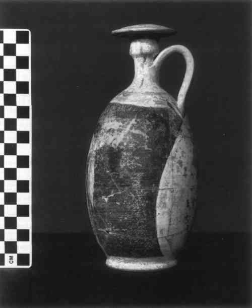
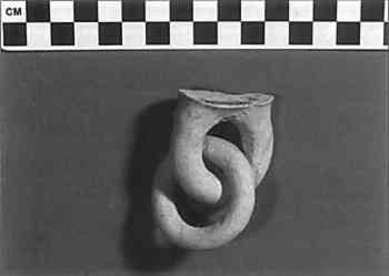
ABSTRACT—Examination of several fragmentary and complete Canosa ceramics in the collection of the J. Paul Getty Museum has been conducted to identify the nature of the pigments employed. Among the colorants identified were rose madder, goethite (FeOOH), Egyptian blue, burnt Sienna, and a carbonaceous black, probably a charcoal black. The range of colors produced on these funeral ceramics is typically white, blue, yellow, pink, light purple, and black, with some of the colorants very susceptible to surface damage because they have not been fired onto the ceramic body. The techniques employed for identification included polarized light microscopy, energy dispersive x-ray fluorescence analysis, Fourier transform infrared microscopy, scanning electron microscopy, x-ray powder diffraction, and optical emission spectroscopy. 1 INTRODUCTIONTHE CANOSA ceramics date from approximately the third to the fourth century B.C. and were produced in southeastern Italy. These ceramics, which were sometimes known as “magenta ware,” were primarily made for burial in tombs and were not intended for everyday use. The ceramic body was usually coated with a white slip before the application of pigment to the slipped clay. Analytical studies of the white slip were carried out by Rinuy and Schweizer (1978), who used scanning electron microscopy, x-ray diffraction, and thermogravimetry in concluding that the white slip was quite pure kaolinite in the hydrated form and that slip was not fired onto the ceramic body. The Canosa ceramics were decorated by applying pigments and by gluing separately molded ceramic components to the main body. Rinuy and co-workers examined this unusual method of construction and identified two different adhesives that had been employed for these ceramic joins (Rinuy et al. 1978; Rinuy and Schweizer 1978). A white adhesive was determined to be a mixture of calcite with most probably an animal glue, while a second, dark adhesive was probably a plant product derived from tree bark. The pigments used on Canosa ware do not appear to have been studied, although there are some notable descriptions of the Canosa ceramics, particularly by Higgins (1956). These pigments were not fired on; some of them are very fugitive, and others may simply be abraded from the ceramic by poor handling. The range of colors on extant examples of Canosa ware is white, pink, yellow, blue, purple, and black. The pink colorant is particularly common, hence the term “magenta ware.” Vases and other decorative Canosa products should be handled carefully. In some cases surface consolidation is required to avoid loss of pigmented surface by handling for study or by physical abrasion during storage. The Canosa ware in the Getty collection is typical of these ceramics. Two examples are shown in figures 1 and 2.
2 EXAMINATION2.1 BLUE PIGMENTALL THE pigment samples studied in this report were mounted in Melt Mount of refractive index 1.662 at 25�C and were used for polarized light examination employing an Orthoplan-Pol polarizing light microscope. Microscopic study of the blue pigment showed the presence of light blue particles of irregular shape, with a refractive index less than 1.66, which are optically anisotropic and exhibit blue-lavender pleochroism. They give a strong pink coloration when a Chelsea filter is inserted into the light path. The Chelsea filter, often used in gemological studies for emerald testing, is useful here for distinguishing between blue pigments by polarized light microscopy (Mactaggart and Mactaggart 1988; Schilling and Scott 1989). These microscopic properties support the identification of this pigment as Egyptian blue. Confirmation of the identification was obtained by x-ray powder diffraction using a Debye-Scherrer camera. 2.2 PINK COLORANTExamination was conducted using polarized light microscopy and long-wave UV excitation of the pigment. A microsample was found to be rose colored, isotropic, with a refractive index less than 1.66. Polarized light microscopy revealed small, poorly defined particles, with an uneven and light red coloration, usually indicative of an organic red pigment. The most common organic red pigment used in antiquity was madder, which usually contains two anthraquinone components, purpurin and alizarin. Purpurin produces a bright yellow-red fluorescence under long-wave UV, while alizarin does not show any fluorescence. The sample did fluoresce, suggesting that the colorant is madder. However, analysis by optical emission spectrophotometry carried out by David McJunkin, a research geochemist at the Laboratory for Historical Colorants, University of California, Los Angeles, provided further information. The plant species usually given as the source of rose madder is Rubia tinctorium L., but no detectable amounts of alizarin were found 2.3 YELLOW PIGMENTSeveral fragmentary pieces of ceramic displayed yellow pigmented surfaces, some of which are very poorly adherent. Yellow surface of the fragment shown in figure 1, for example, had already become partially detached by storage in a polyethylene bag. The pigment had been attracted from the ceramic surface to the bag itself by electrostatic forces. Polarized light microscopy showed that the pigment consisted of small yellow-brown clumps with yellow rod-shaped particles whose refractive index is difficult to determine. Most standard samples of yellow ochre only contain incidental needle-shaped crystals, but the sample from the ceramic consisted mostly of these fine crystals. The particles are anisotropic and appear very similar to one of the yellow ochres in the Edward J. Forbes collection, labeled as 3.04.11, Naples 1919. From a collection of 10 different samples, only this yellow ochre sample proved to be similar. A microsample of the pigment from the ceramic fragment was added to a pellet of packed potassium bromide powder and analyzed by Fourier transform infrared spectroscopy in the transmission mode by Michele Derrick (1988). A very close match to the spectrum acquired was given by a reference spectrum of geothite, α-FeOOH. Identification of the ochres in terms of the precise isomer of the iron oxyhydroxides is often difficult, but here relatively pure goethite appears to be the source of the yellow pigment used. 2.4 BLACK PIGMENTX-ray fluorescence was used first to examine the black pigment areas in situ. It was not possible to demonstrate that the black areas contained higher iron or manganese content than the red body of the ceramic itself. The logical conclusion is that the black surface is likely to be an organic black pigment rather than an iron black. Caution must be exercised in coming to general conclusions too quickly, however, since a sample of the black pigment examined by polarized light microscopy showed the clear presence of an opaque black mixed with an iron oxide pigment. Examination showed that the iron oxide pigment did not contain any manganese, an impurity usually associated with umbers. The red pigment which was mixed with the black charcoal is also very similar in optical properties to a McCrone reference sample of burnt Sienna rather than an umber or an ochre. The black surface was examined further in situ with x-ray fluorescence spectroscopy using a sulfur secondary target with helium flushing. The advantage of this method of analysis in the case of carbon blacks is that excitation of phosphorus is greatly enhanced by the use of a sulfur secondary target ensuring that phosphorus will be detected with good sensitivity even at 0.1% by weight. No phosphorus was detected in the black pigment, which implies that bone black or ivory black were not employed; this fact, together with the polarized light microscopy study, indicates the use of a carbon black from a plant source. The use of mixtures of natural earth ochres with carbon to create Examination of the pigmented surface by scanning electron microscopy was carried out on a small flake detached from the ceramic. Analysis by wavelength dispersive methods showed that the pigment particles are principally carbon and occur in small clumps; many of the particles are less than 1 micron in size. A scanning electron micrograph showing part of the topography of the black surface revealed surface structure typically associated with carbon black pigment. 2.5 WHITE SLIPFourier transform infrared spectroscopy confirmed the results for this slip obtained by Rinuy and Schweizer (1978), namely that the material is a relatively pure kaolinite. This part of the study was not taken any further because of the work that has already been reported. 3 CONCLUSIONSTHE WORK presented here has identified the nature of the pigments on samples of Canosa ware in the collection of the J. Paul Getty Museum. Some of the pigments from these vases are very fragile and may require surface consolidation to ensure their stability. The range of pigments used is quite typical for the period of manufacture in the early centuries B.C., although it is unusual to see rose madder used as ceramic colorant; this material would burn out in firing and so be lost in a fired ceramic surface. ACKNOWLEDGEMENTSTHE AUTHORS wish to thank the following for essential advice and guidance: Michele Derrick, associate scientist, Analytical Section, Getty Conservation Institute (GCI), for carrying out the Fourier transform infrared examination; Eric Doehne, associate scientist, GCI, for electron microprobe examination of one sample; Ken Hamma, associate curator, Department of Antiquities, J. Paul Getty Museum; Max Saltzman, scientific consultant, GCI; David McJunkin, research geochemist, Laboratory for Historical Colorants, University of California, Los Angeles; and Janet Boley, part-time scientific assistant, for carrying out x-ray diffraction analysis at the Los Angeles County Museum of Art. REFERENCESDerrick, M.1988. Infrared analysis report on the Canosa pigments. Internal Getty Conservation Institute report. Unpublished. Higgins, R.1956. Magenta ware. The British Museum Yearbook. 1–32. Mactaggart, P., and A.Mactaggart. 1988. A pigment microscopist's notebook. Unpublished. Profi, S., L.Weier, and S. E.Filippakis. 1974. X-ray analysis of Greek Bronze Age pigments from Mycenae. Studies in Conservation19:105–112. Rinuy, A., and F.Schweizer. 1978. Analysis of the white “ground” and ancient adhesives found on Canosa vases (south Italy) of the third century B.C.Proceedings of the 19th International Symposium on Archaeometry and Archaeological Prospection: Bonn. 14–17.
Rinuy, A., F. van derWielen, P.Hartmann, and F.Schweizer. 1978. Ceramic insolite de I'Italie du sud: Les vases hellenistiques de Canosa. Extrait de Mus�e d'Art et d' Histoire: Geneva. N.s. 26. 141–69. Schilling, M. P., and D. A.Scott. 1989. Letter to the editor. Journal of the American Institute for Conservation28:137. AUTHOR INFORMATIONDAVID A. SCOTT has been head of the Museum Services Section of the Scientific Program at the Getty Conservation Institute since 1987. He has been a lecturer in conservation at the Institute of Archaeology, University of London, Department of Archaeological Conservation and Materials Science, and, since 1984, an editor of Studies in Conservation. He was named a fellow of the International Institute for Conservation in 1989. His principal research interests are the analysis and technical study of ancient metallic objects and their corrosion products, the conservation of metallic artifacts, the study of Chumash Indian rock art, and the archaeometallurgy of ancient South America, particularly Colombia and Ecuador. Address: Getty Conservation Institute, 4503 Glencoe Avenue, Marina del Rey, Calif. 90292–6537. Michael Schilling is associate scientist in the Analytical Section and Museum Services Section of the Scientific Program at the Getty Conservation Institute. His principal research interests include methods and application of thermal analysis and the examination and analysis of painted surfaces. Address: Getty Conservation Institute, 4503 Glencoe Avenue, Marina del Rey, Calif. 90292–6537.
 Section Index Section Index |