THE USE OF DIGITAL IMAGE PROCESSING TO CLARIFY THE RADIOGRAPHY OF UNDERPAINTINGJames R. Druzik, David L. Glackin, Donald L. Lynn, & Raim Quiros
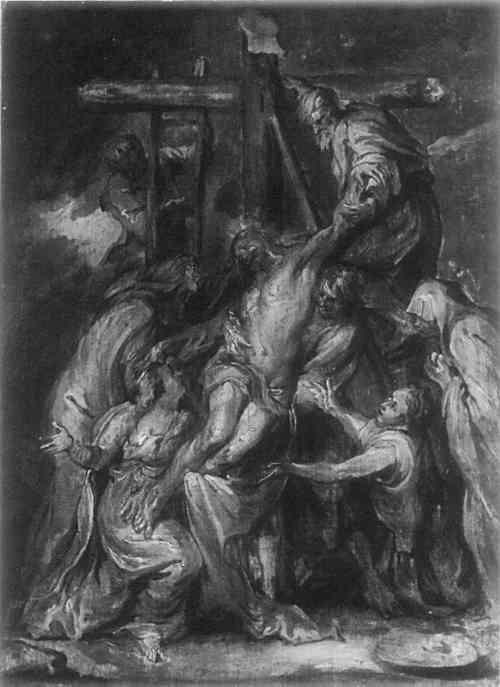
ABSTRACT—Digital Image Processing is a computer-assisted technique which can help the conservator interpret the visual information contained in the many photographic images produced when documenting a work's condition. In this paper some of these techniques are utilized to assist in evaluating the radiography of a complex 17th century panel painting. 1 INTRODUCTIONTHE PURPOSE OF digital image processing is threefold; to improve the appearance of an image to a human observer, to extract from an image quantitative information that is not readily apparent to the eye, and to calibrate an image in photometric or geometric terms. Image processing is an art as well as a science. It is a multidisciplinary field which contains elements of photography, computer technology, optics, electronics, and mathematics. Image processing was first developed largely in response to the needs of the space program. The Jet Propulsion Laboratory (JPL) of the California Institute of Technology began working in the field over twenty years ago. In 1965 JPL established the Image Processing Laboratory (IPL)1 in support of NASA's unmanned spacecraft exploration of the moon and planets. But in recent years, these techniques have found wide application outside this initial application. Medical science has applied digital image processing; Landsat images have led to more efficient oil exploration. mining, improved forest management, and the measurement of broad scale insect infestation. A newly developed area of application is in conservation research and condition documentation, an example of which is the study at hand. 2 IMAGE PROCESSING OF WORKS OF ART AT IPL2.1 ScanningThe first procedure, called scanning, converts the image of an artwork on a film negative to a computer-readable form. Of the several machines in the IPL that can perform this function, the PDS Microdensitometer was used because it is the most accurate. The negative is placed on a glass transport through which passes a narrow beam of light whose brightness is read by a detector. The stage is movable and is under computer control. The negative is scanned by commanding the transport to move, momentarily stopping at each of a number of closely spaced grid points to allow the density of the negative to be read. The scanning process divides the image into a two-dimensional array of data samples or pixels (short for ‘picture elements’) which represent image brightness (grey level). In the typical ‘8-bit’ encoding scheme, there are 256 possible grey levels, ranging from 0 (black) to 255 (white). The eye can only The scanned image is stored on computer tape. The typical image in this study had roughly 1000 pixels vertically by 1000 pixels horizontally, so such an image is represented by one million image samples. After scanning the image is processed on an IBM 370/158 computer with the aid of the disc drives which act as temporary storage during processing. 2.2 Digital Image ProcessingDigital image processing consists of the application of a series of computer programs to an image to produce a desired result. The image processing computer language in the IPL, VICAR (Video Information Communication and Retrieval),1 is a command-oriented computer language library of parameterized application programs. Complex image manipulations can be executed with the use of a single command. VICAR was designed for ease of use, and does not require intimate knowledge of the machine. Over 300 VICAR applications programs are currently available. 2.3 Playing BackAfter an image is processed, it is often converted back into the form of a photograph, a process known as ‘playing back.’ The playback device used in this study, Dicomed, read the pixel grey levels in the processed image and used them to adjust the intensity of a twenty-micron flying electron spot. The spot scans the film negative, stopping momentarily at the location of each pixel and exposing the film to the proper level. The negative is developed and black-and-white prints produced in the usual manner. For a technical description of IPL hardware the reader is asked to refer to authors. 3 APPLICATION OF IMAGE PROCESSING TO PAINTING CONSERVATIONFIGURE 1 shows a grisaille painting, “The Deposition” formerly attributed to Van Dyck. Jan Erasmus Quellinus (1634–1715) has been suggested;2 in any case it is technically attributable to the period. Conservation treatment records indicate that in 1975, mass spectrometric analysis of a sample of white and red pigment indicated basic lead carbonate and mercuric sulphide.
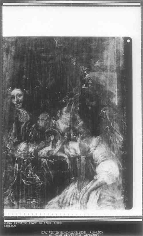
The x-ray radiography revealed that there are two painting structures (Fig. 2); the lower one on wood panel, appears to be a sitting room scene. “The Deposition” is painted on paper (100% linen fiber by microscopic examination) which has been adhered over the lower image. Two objectives necessary to restore the confused x-ray image were defined: first, to remove or suppress the wood grain pattern interference, and secondly, to separate the two images, isolating each one digitally and photographically. In order to try this strategy one additional x-ray technique was utilized to give the IPL more raw data from which to work; that is a secondary electron emission radiograph.
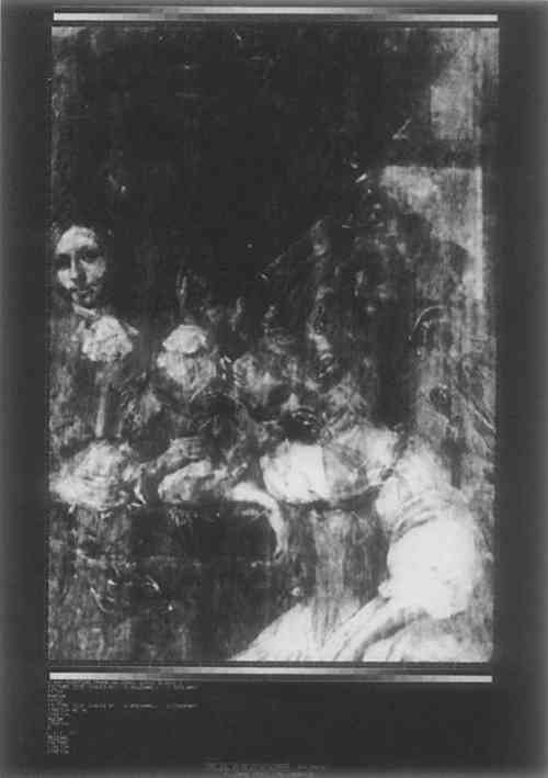
3.1 Removal of the Wood Grain Striations from the X-ray ImageThe oak panel support created a series of striations in the x-ray image. Figure 2 is a contrast-enhanced version of this image. The striations are roughly straight, roughly vertical, and evenly spaced. If they were precisely so, they would be relatively The first method tried, which did not work, involved using a median filter one pixel high and a few pixels (5–9) wide. The second method, which produced excellent results, is a two-dimensional Fast Fourier Transform. This method produced a photographic plot which shows the size distribution of features in the original image. The wood grain was the only feature being periodic; thus with this data a mathematical filter could be constructed which removed this high frequency pattern. Figure 3 shows the original x-ray, filtered and relatively free of vertical striation.
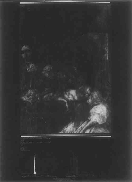
3.2 The Subtraction of the Two Superimposed ImagesThe subtraction of the two superimposed images was accomplished by means of a suitable sequence of grey-level matching, spatial filtering, subtracting and averaging. Two intermediate products were produced from the x-ray emission image. To make the first intermediate product, the x-ray emission image was subjected to a high-pass spatial filter, to enhance feature edges and fine detail. The distribution of grey levels in the resulting image was approximately matched to that in Figure 3. The resulting image is the first intermediate product. To make the second intermediate product, the same steps were followed, except that a low-pass spatial filter was used. The final product, Figure 4, the subtraction of the superimposed images, was made by averaging the two intermediate products.
4 DISCUSSIONWHAT THIS ASPECT of the study generated was a series of images which taken alone yield some, but not a great deal of new information compared to what was present in the original material. There are three features in Figure 4 which are obscure in the original x-ray, particularly when displayed on a high resolution computer monitor. These features are the face of the seated woman, the left hand of the man in the left part of the painting, and what may be a person looking in through the window. After these features are pointed out, they can be found with difficulty in Figure 2, but they required the processed image to find them initially. The hand stands out better after the removal of the wood grain striations, because the striations were parallel to the fingers. The face stands out better after removal of the over-image. Digital image processing does not create information which is not contained in the original images in some form. What is achieved, rather, is the isolation of discrete units of visual information with a common numerical “denominator.” Taken as a block of visual images and information, and aided by a knowledge of the processing steps, the conservator is led through the many levels of visual data step by step to clarify the relative contributions of both images and, perhaps, to give a little more insight into the structure and state of completion of the under-image. It should be kept in mind that this was a very complex example, as several factors interfered to make this study more difficult than what might be expected with other Northern European paintings. In particular, the fact that a large amount of lead white occurs in the regions where we had hoped to gain the most insight into the lower painting created an unfortunate and unanticipated complication. ACKNOWLEDGEMENTSTHE AUTHORS would like to acknowledge the patience, encouragement and continued support of William Liesher, Head of Conservation, and Dr. Pieter Meyers, Senior This work was supported under the Cal Tech President's Fund # PF-183. This paper presents one phase of research conducted at the Jet Propulsion Laboratory/California Institute of Technology, under NAS 7–100, Aeronautics and Space Administration. REFERENCESCastleman, Kenneth R., Digital Image Processing (1979), Prentice-Hall Inc., p. 383. Personal communication. Scott Schaeffer, Curator of European Paintings, Los Angeles County Museum of Art.
 Section Index Section Index |