The Tale of the Red-winged Blackbird: A Case Study of Varnish Removal from a Watercolor Painting
by Pamela J. YoungIntroduction
The exhibition Mark Catesby's Natural History of America: the Watercolors from the Royal Library, Windsor Castle came to a close at the Dewitt Wallace Gallery in February, 1998, while the paper lab prepared works on paper from the permanent collection for a replacement exhibit called Drawing on Nature. the staff of the paper lab then included Mary Studt, a Getty advanced intern, and Amy Gerbracht, a graduate intern. in the midst of feverish activity, Margaret Pritchard, curator of prints, maps and wallpaper, came to the paper lab with eleven watercolors depicting North American birds. They were offered for sale, unattributed but clearly in the style of several, well-known eighteenth-century European artist-naturalists represented in the Foundation's collection, among them Catesby, George Edwards, Eleazar Albin, and Georg Ehret. the provenance indicated that the eleven were originally part of a much larger group owned by John Drayton (1713-1779) of Charleston, South Carolina, a colonist of considerable stature and social position. Two paintings held immediate interest for the curator because they appeared to be direct copies of prints included in the first set of Mark Catesby's Natural History of Florida, Carolina and Bahama Islands, 1732-1747, a seminal volume in the study of flora and fauna of the American British colonies. They were the Whip-or-Will or Lesser Goat Sucker and the Red-winged Blackbird. Could these be previously unknown Catesby works? Both curator and dealer were intensely interested, for it had a direct bearing on the price.
The initial examination revealed two distinct types of paper, each with an obvious mold pattern and many with at least a portion of a watermark. Both patterns were reproduced by a simple tracing on polyester film so that the curator could take to them to England for comparison with similar paintings in the Sloane Collection at the British Library. Sir Hans Sloane, a serious collector of natural history illustrations and specimens during the first half of the eighteenth century, had wisely inscribed title and artist on many paintings as they entered his collection. in fact, when the Mylar facsimiles were compared with paper supports in this collection, both mold patterns and watermarks matched exactly those attributed to George Edwards. Consideration of both materials and technique used by the watercolorist confirmed that the English naturalist George Edwards had painted all eleven.

Fig. 1. George Edwards. the Red-winged Blackbird. Watercolor on paper. Colonial Williamsburg Foundation. Gift of Sumpter Priddy, 1997. the painting was initially attributed to Mark Catesby.
The blackbird in particular caught the eye of the conservators (fig. 1). It was unique in that a dark brown, severely crackled varnish layer covered the body and wings of the bird. in fact, the varnish was so disfiguring that the dealer had decided to make a gift of the Red-winged Blackbird to the Foundation, assuming that the new paper lab might be able to provide the extensive treatment required. Through analysis, conservation, and restorative treatment, both curator and conservators hoped to support the identity of the artist and return the appearance of the painting to a state closer to the artist's original intent and make it worthy of display.
The painting in question is executed in watercolor and gouache with graphite underdrawing. the support is a handmade antique-laid paper, slightly textured with obvious sizing, of medium thickness and composed primarily of cotton and linen fibers. We estimated the original paper color had been off-white but had aged to beige/brown.1 at this point it was difficult to estimate which pigments had been employed due to the color shift imposed by the varnish layer.
Regarding the varnish, several factors suggested that the coating was not original to the painting. to the curator's knowledge, no other watercolors by Edwards are coated in this manner, for example, the Razor-billed Black-bird of Jamaica, an image also rendered in black pigment in the collection of Windsor Castle. the coating had been haphazardly applied, at times overlapping onto and causing discoloration of exposed paper. Because the bird is painted in an otherwise precise manner, as seen from infrared photographs, it seemed unlikely that the artist would have applied the final coating rather carelessly. the coating resembled a darkened accretion on the verso of the painting, possibly the residue of a substance used to adhere the painting into an album. This was a common practice in the eighteenth century. It seemed plausible then that the individual who had stuck the painting into an album had applied the coating as well.
A Brief History Of Varnish on Works on Paper
A cursory search of both contemporary and historical literature produced little information on the use of varnish on paper-based artifacts. As known from experience and conversation with other conservators, old varnish layers appear on utilitarian objects such as maps, globes, mariner's quadrants, and decorative objects, including prints, wallpaper, fraktur and folk art paintings, and photographs. Varnish coatings functioned as barriers to moisture, fly specks, and surface abrasion. They were applied to imitate the appearance of oil paintings, to saturate dark pigments, and add detail with high gloss. Traditional materials used as both binder and varnish include gum arabic, cherry gum (a generic term for the exudite of cherry, apricot, and almond trees), gum tragacanth, and gum accroides. Shellac was used in the eighteenth century to varnish large framed prints in place of glazing, at a time when manufactured glass was very expensive and the size was limited.
According to Painting Materials, a Short Encyclopedia, by Gettens and Stout, the use of plant gums as sizing and medium is mentioned as early as the twelfth century.2 Gums are a group of non-crystalline, structureless materials, composed mainly of carbon, hydrogen, and oxygen and differ from protein binders and adhesives in that gums contain practically no nitrogen.3 Characteristically gums swell in water, and when completely dissolved, form a clear, viscous solution referred to as mucilage. They are insoluble in alcohol. C. V. Horie, in Materials for Conservation, describes plant gums as high molecular weight polysaccharides, which degrade by hydrolysis and photo-oxidation, much like starch.4 Ralph Mayer's a Dictionary of Art Terms & Techniques includes this note on preparation: "When a solution of gum arabic is heated to the boiling point its character changes: it becomes dark, acquires a pronounced odor, and will not function properly in the recipes calling for a hot water solution; however, it may be used as a paper adhesive."5
Condition

Fig. 2. Quilted distortion caused by varnish layer. Verso, raking light
The blackbird painting was generally in poor condition. the turbid coating had caused major instability of both design layer and support, as well as a serious compromise of aesthetic quality. Mechanical damage to the paper support included overall planar distortion and a quilted effect under most of the coated region (fig. 2). Drying and contraction of the coating had caused the media to crack and cup, pulling the uppermost layer of paper fibers with it (fig. 3). the paint layer was obviously discolored by penetration of the brown coating and originally may have resembled the grayish black of the bird's legs.
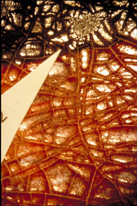
Fig.3. Detail, crackled varnish with marker indicating location of sample removal.
The verso was moderately discolored overall with a pronounced mottled effect apparently caused by mold growth. Numerous, large metallic inclusions in the paper were the vivid green of corroded copper and were surrounded by darkened, degraded paper.
Before proceeding with treatment, the conservators and curator asked several essential questions: Did the artist apply the varnish? Why was it applied and what was it composed of? Could the varnish be removed without damage to the watercolor below? Using various analytical techniques, answers to these questions began to unfold during further study of the painting.
Analysis

Fig. 4. IR reflectagraph image: a. overall
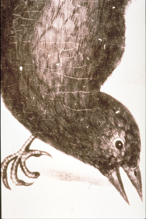
Fig. 4. IR reflectagraph image: b. detail.
With the aid of infrared radiation, an IR-vidicon camera, and a monitor, we could see a highly detailed rendering beneath the coating and confirmed the presence of a carbon black pigment (figs. 4.a-b). the remarkable image also provided the motivation to commit what we knew would be a significant investment of time and energy to the project. This truer image of the blackbird was documented as a photographic print as well. Infrared photography enhanced the presence of inclusions in the coating, abrasion and cracking of the paint layer and revealed distinct boundaries between the black portion of the wings and the yellow/orange epaulets.

Fig. 5. Epaulets revealed by x-radiography.
The results of Grenz radiography suggested that X-ray dense pigments were present in the brightly colored regions of the wings (fig. 5). Lead-based pigments such as minium (red lead, tetroxide of lead), massicot, and litharge (yellow monoxides of lead), lead-tin yellow, and the mercury-based pigment vermilion may be present.
| SAMPLE | SHORT WAVE UV (254 nm) | LONG WAVE UV (366 nm) |
| known - gelatin | grey, low brightness | bright white, high brightness |
| known - gum arabic | pale yellow, medium brightness | bright white, high brightness |
| unknown - coating over the red portion of win | pale yellow, low brightness | orange, high brightness |
| unknown - coating over the black portion of the body | warm brown, low brightness | yellow brown, medium brightness |
| accretion on lower border of the paper verso | warm brown, low brightness | yellow brown, medium brightness |
The painting was observed overall under ultraviolet light to examine fluorescence of the coating. Descriptions of fluorescence were organized in a table for comparison; the results were inconclusive (fig. 6).
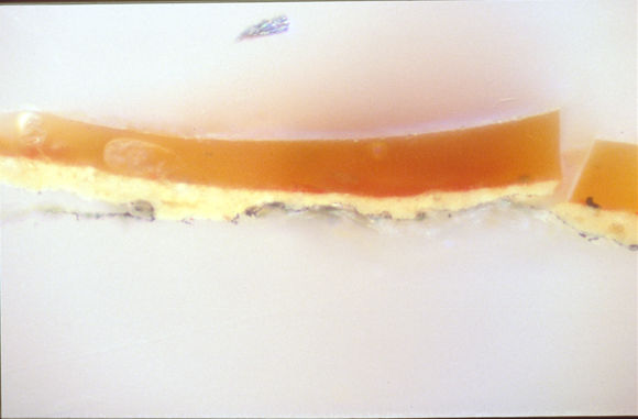
Fig. 7. Cross-section sample, epaulet region, 50X, daylight; varnish layer/red watercolor pigment/yellow watercolor pigment/graphite/paper fibers.
Micro-samples were taken from the epaulet and body regions and mounted in plastic resin for cross-sectional viewing. Visual analysis indicated varnish and paint were discreet layers (fig. 7). We stained the samples with triphenyl tetrazolium chloride and fluorescamine to determine the presence of either protein or carbohydrate.6 When stained with TTC the varnish layer became brown, indicating the presence of a carbohydrate. Fluorescence of the cross-sections when stained with fluorescamine was not intense, suggesting that little or no protein was present.
Samples of the coating from the body and the epaulets, and the brown accretion from the verso, were tested with ninhydrin to determine whether protein was present in the samples.7 We compared the ninhydrin test papers with control papers and filter papers immersed in a .5% gelatin solution. Sample papers immersed in solutions of the coating did not show the dark purple color one would expected if protein were present in the coating layer. Testing suggested that protein was not present in any of the unknown samples.
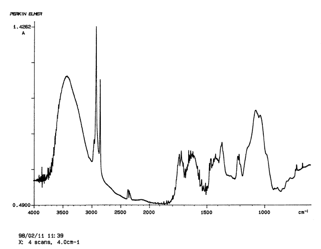
Fig. 8. Infrared spectrograph of varnish sample.
Infrared spectrometry gave us irrefutable evidence that the varnish was indeed gum arabic (fig. 8). Samples of the coating covering the body and the epaulets, and the brown accretion from the verso were taken and crushed into a powder. These were examined by Fourier transform infrared spectrometry and the sample spectra compared with those of known protein-based and carbohydrate-based samples.8 in each instance the spectra confirmed the sample as a gum arabic.
Treatment
| Sample are | H2O | NH4OH solution phH 9.0 | H2O : i-propanol 1:1 | i-propanol | acetone : i-propanol 1:1 | acetone | H2O acetone 1:1 | mineral spirits |
| red wing | 2 | 5 | 1 | 1 | 1 | 1 | 1 | 1 |
| black body | 5 | 5 | 3 | 2 | 2 | 2 | 3 | 1 |
To completely convince ourselves that the gum layer could be removed safely, we conducted a series of solvent tests on the coating with mineral spirits, isopropanol, acetone and water, outlined in another table (fig. 9). Water quickly swelled and dissolved the coating in the area of black watercolor. Isopropanol and acetone only slightly dissolved the coating in the same areas. Mineral spirits had no discernible effect. the coating was less water soluble in colored areas, where it was slightly swelled by water alone. We speculated that interaction with the metal-based pigments may have affected drying or aging of the varnish layer, a theory supported by C. V Horie, who notes in his description of plant gums that their molecules can be cross-linked by trivalent metal ions, e.g. lead and mercury salts.9 Further, the crackle pattern was finer and more extensive in the black pigment areas, perhaps due to the cumulative effect of a consistently higher temperature in this region caused by the absorption of IR energy. the Wolbers cleaning systems were reviewed to see if one might be applied successfully. a variety of moisture delivery systems were considered including agar and Gore-Tex, but methylcellulose seemed to have the greatest potential for providing the appropriate moisture release rate. the addition of ammonium hydroxide to water slightly increased the solubility of the coating in both regions. a combination of water and isopropanol caused the coating to become soft and coagulated, and it was this reaction that allowed the greatest control in manipulating the coating without significant alteration of the watercolor layer beneath.
Both curator and conservators decided that the benefits of varnish removal outweighed the risks, and that the treatment should be undertaken to reduce stress on the paper and paint layers and reveal detail obscured by the dark coating. the removal procedure involved softening and swelling the gum with a poultice followed by mechanical action to reduce the excess. Although the removal process required minimal tools and supplies, the success was all in the technique. Achieving the desired result, that is, removal of the varnish without significant alteration of the paint layer, required extraordinary skill and sensitivity to the "moment of truth," when all the variables were right for successful execution of the procedure.
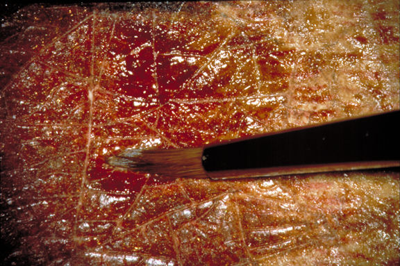
Fig. 10a. Process of varnish removal: application of poultice.
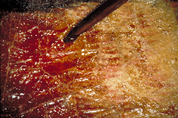
Fig. 10b. Process of varnish removal: mechanical removal of swelled varnish.
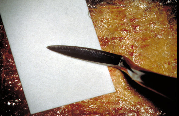
Fig. 10c. Process of varnish removal: application of blotter.
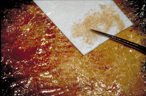
Fig. 10d. Process of varnish removal: adsorption of varnish residue.
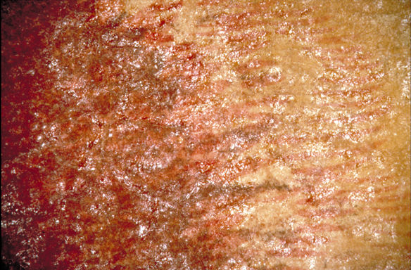
Fig. 10e. Process of varnish removal: after treatment.
After extensive testing, the coating was removed overall by applying poultices of methylcellulose with the addition of isopropanol (3:1) and ammonium hydroxide to a pH of 8.0 (figs. 10a-e). the addition of alcohol to the methylcellulose caused less swelling of the paper. Both poultice material and coating were removed with a micro-spatula, swabs, and blotter triangles, working under magnification. Progress was slow, the removal proceeding at the rate of one square centimeter per half-hour in the black painted area, and a square centimeter per hour in the colored epaulets. Each treated area was dried under lightly weighted layers of Hollytex and blotter to keep further distortion and cracking to a minimum. an 8" x 10" print of the IR image provided an important reference for locating thinner and thicker areas of the paint layer. Raking light examination of the verso showed significant reduction of distortion in the treated area. an unavoidable side effect was formation of tide lines; these would be removed during subsequent washing.

Fig. 11. During treatment, partial removal of varnish.
The varnish removal became a three-phase process. the first phase involved minimal moisture application with intrusive, direct mechanical action to remove the bulk of the coating, but leaving a protective residue over the paint layer (fig.11). This step relieved the mechanical stress imposed by the thickness of the desiccated, shrunken varnish on both design layer and support. the second phase included moderate moisture application and moderate mechanical action in dual forms of dampened swabs rolled through Japanese paper and water sprayed during the blotter wash, using blotter strips and more Japanese paper to lift varnish and methylcellulose residues and cotton fibers. As the watercolor layer was exposed and became increasingly sensitive, the suction table was employed to allow maximum moisture application and minimum surface manipulation. This constituted the third phase.
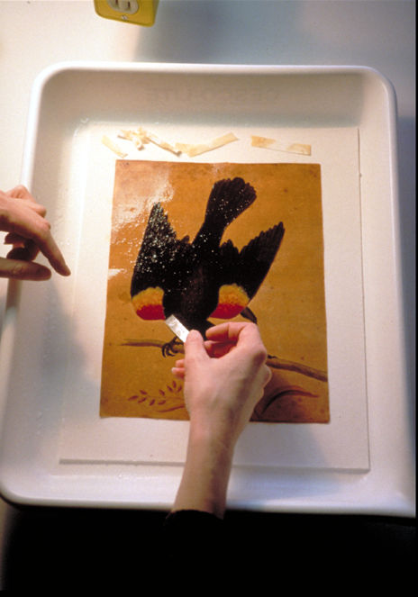
Fig. 12. Washing the watercolor on a blotter stack and blotting varnish residue from the paint surface.
Residual gum and methylcellulose remained on the surface, visible as glossy, whitish patches. the painting was washed on a blotter stack to allow maximum wetting, with significant removal of discoloration from both the paper and paint (fig. 12). As we sprayed the object, the varnish residue and poultice swelled and were blotted away. We removed numerous cotton wool, blotter, and Japanese paper fibers with tweezers. Washing continued on the suction table with ammoniated water followed by a calcium hydroxide/water solution with continued blotting. a magnesium bicarbonate solution was applied to areas where metallic inclusions had been excavated.
Localized bleaching with a 3% hydrogen peroxide solution reduced discoloration over the body of the bird and in the background. Light bleaching for 2.5 hours lightened the discolored organic coating and enhanced the contrast in the black painted portion.
We resized the recto and verso surfaces with a dilute gelatin solution, which added body to the paper overall, reduced the fissure size in paint cracks, and slightly saturated and isolated the original watercolor.
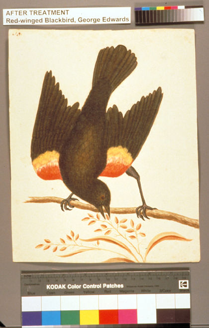
Fig. 13a. After treatment: overall.
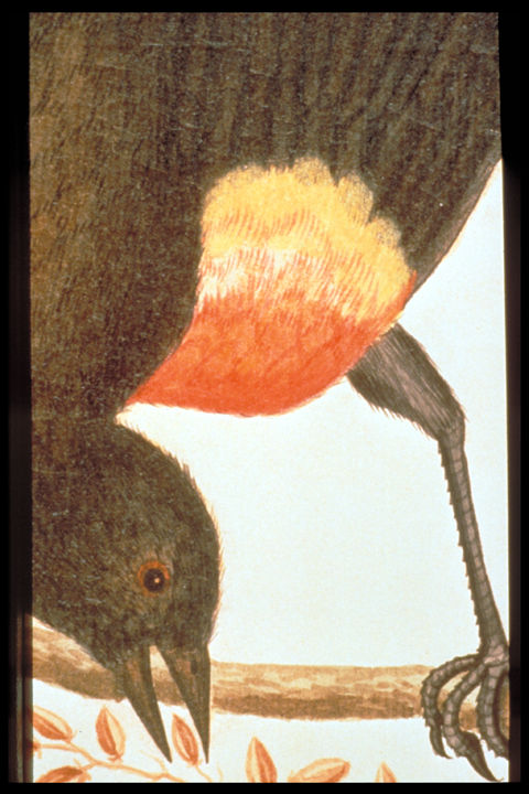
Fig. 13b. After treatment: detail.
Japanese tissue strips reinforced sharp creases and weaknesses in the support and helped keep them in plane during the final flattening. Very thin antique paper patches camouflaged severe areas of discoloration caused by metallic inclusions. Losses in the paint layer were inpainted with watercolor paints (figs. 13a-b).
Conclusion
The entire project consumed over 250 hours, and multiple, unique opportunities presented themselves along the way. We collaborated with the curator on confirming the attribution. We provided a highly successful treatment for an object deemed too disfigured for sale or exhibit. the interns undertook a complex and multi-faceted treatment and got experience designing a related exhibit. in an effort to promote the benefits of conservation/curatorial collaboration, we mounted a small display adjacent to the primary exhibit, Drawing on Nature. Text panels described the process of analysis and treatment; case bonnets below displayed tools and supplies. an enormous graphic on the opposite wall represented the requisite dos and don'ts of paper preservation.
Acknowledgments
Mary Studt was principally responsible for analysis and treatment of the Red-winged Blackbird painting. I would like to thank Colonial Williamsburg staff for their assistance with documentation of the project and Dr. James Rice at the College of William and Mary for use of the Fourier transform infrared spectrometer.
Notes
1. Classifications made by comparison with Print Council of America, Paper sample book, 1996.
2. Rutherford J. Gettens and George L. Stout, Paintings materials, a short encyclopedia (New York: Dover Publications, Inc., 1966), p. 28.
3. Gettens and Stout, Paintings materials, p. 28.
4. C. V. Horie, Materials for conservation, organic consolidants, adhesives and coatings, (Oxford: Butterworth-Heinemann, 1996), p. 141.
5. Ralph Mayer, a dictionary of art terms & techniques (New York: Thomas Y. Cromwell, 1975), p. 179.
6. Richard Wolbers et al. Notes for workshop on new methods in cleaning paintings (Los Angeles: Getty Conservation Institute, 1990) p. 60-61.
7. B. L. Browning, Analysis of paper (New York: Marcel Dekker, Inc., 1969), p. 105.
8. Amy Snodgrass and Beth A. Price. the Gettens collection of aged materials of the artist: FT-IR spectral library and catalogue of the raw materials (Strauss Center for Conservation and Philadelphia Museum of Art, 1993), pp. 100.B03-06, B-08, B-12, B13, B15, B16, B17, B20, B-22, 100.B31, B36, B37, B50, B46, B47.
9. Horie, Materials for conservation, p. 141.
Pamela J. YoungConservator of Works on Paper
Colonial Williamsburg Foundation
Publication History
Received: Fall 1999
Paper delivered at the Book and Paper specialty group session, AIC 27th Annual Meeting, June 8-13, 1999, St. Louis, Missouri.
Papers for the specialty group session are selected by committee, based on abstracts and there has been no further peer review. Papers are received by the compiler in the Fall following the meeting and the author is welcome to make revisions, minor or major.