Photographic Science and Engineering. Vol. 28, Num. 4, July/August 1984. p.166-71
Received May 4, 1984.
© 1984, Society of Photographic Scientists and Engineers.
The image structure and changes in image structure during deterioration of albumen prints, a silver printing-out paper extensively used in the 19th century, were investigated by use of transmission electron microscopy. Albumen print images are composed of roughly spherical particles with diameters ranging from <3 to ~50 nm, with most between 5 and 20 nm. The image material is concentrated in the upper half of the albumen layer. Various influences on image formation are discussed, including gold toning, which distorts the original particle into an elongated, irregular shape. Because of the photolytic mechanism of image formation, low-density areas have fewer and smaller particles than high-density areas, leading to rapid highlight density loss through an oxidative-reductive deterioration process. The image structure and resistance to deterioration of albumen prints and a typical developing-out paper are compared. In high-density areas of albumen prints incubated at high RH, the image deposit is completely re-formed, underscoring the need to control the relative humidity of the storage areas for these objects. A means of predicting micro-structural changes based on initial particle size, size frequency distribution, and spatial distribution is presented.
Because of the dominance of the albumen print material in photographic practice from '4855 to 1895, a substantial portion of the legacy of 19th-century photography exists in this form. The nature and structure of these prints, and particularly their image stability, are a concern to archives, libraries, and museums. Most albumen prints have deteriorated, showing overall density loss, severe highlight density loss, and a marked shift in image hue. A recent investigation' found thiosulfate retention from improper processing unlikely to be the explanation for such widespread deterioration; thiosulfate was adequately removed fairly rapidly, and even well-processed images were unstable in moist air at moderate temperatures. These results, together with the evidence from >100 years of natural aging, suggested that more fundamental factors, perhaps related to image structure, were responsible for the deterioration.
A relationship between silver image structure and resistance to sulfiding and oxidative-reduction deterioration has long been known. Baekeland in 18972 related the poor image stability of printing-out to a much smaller particle size, though he still had doubts about whether the image was composed entirely of metallic silver. He also noted that among developed images, coarse-grained prints were more resistant to attack than fine-grained ones. Considerable progress has been made in our understanding of the mechanisms of silver image deterioration and the relationship between image structure and resistance to attack. The relevant structural features are much too small to be observed with an optical microscope and can be studied only by use of electron microscopy. Our study of albumen-print image structure has the dual objectives of investigating image formation in albumen prints and relating characteristic structural features to deterioration patterns observed in both natural aging of historical prints and the incubation of prints made in the laboratory.
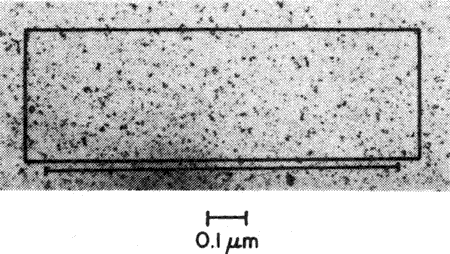
The albumen print material3-5 consisted of a relatively thin paper support coated on one side with an image-bearing layer of hen's egg white. Overall sheet thickness was typically ~0.1 mm, and the albumen layer varied in thickness up to ca. 12 ìm. The albumen coating (containing 1-2% ammonium or sodium chloride) was applied by floating individual sheets on a tray of albumen solution; they were then hung vertically to dry. This caused the albumen layer thickness to vary from top to bottom. A second layer was often applied and the sheet hung upside down during drying to compensate for the uneven first coating. To achieve a net gain in coating thickness, some method for lowering the solubility of the first coating was required. Bathing the coating in low-molecular-weight alcohol solutions was frequently mentioned in the literature but apparently not widely used commercially. Since the solubility of dried albumen declines naturally over a few months, most manufacturers simply "cured" the product between coatings. In our study, when the first coating of laboratory-made prints was coagulated in a 70% solution of 2-propanol, a definite interface between the layers was visible in the electron micro-graph cross-sections. Only one 19th-century print among the eleven examined had such an interface.
For use, the albumen paper was sensitized by floating it on a 6-10% silver nitrate solution. A more sensitive paper with better keeping properties was obtained when up to 5% citric acid was added to the silver solution. The albumen print image was produced by printing-out, i.e., entirely by photolysis. Processing included an initial wash to remove all soluble silver compounds, gold toning, fixation in sodium thiosulfate, and a final wash.
Figure 1. Cross-section electron micrograph of a 19th-century albumen print, showing albumen layer, image deposit (concentrated in top half of albumen layer), and uppermost paper fibers (spongy area at bottom) (20,000x).
Figure 2. Image deposit from Fig. 1 at higher magnification (80,000X).
For studies of image structure and the various influences on image formation and deterioration, cross-section electron micrographs cut normal to the print surface were prepared. In all, 11 different 19th-century prints and ca. 40 laboratory-made prints were examined, usually at several density levels. Typical electron micrographs at low and high magnification of a 19th-century albumen print (ca. 1890) are shown in Figs. 1 and 2.
Two features are noteworthy: The image material is largely confined to the upper half of the albumen layer, and the particle size and number of particles progressively decrease at greater depth in the coating. All printing-out papers share these features, and the presence of a vertical gradient of density (which can be clearly seen at the lower magnifications possible with the optical microscope6) is a useful characteristic in differentiating printing-out papers from developing-out papers. The progressive decrease in particle size and number is due in part to the shielding effect of photolyzed silver, which forms initially in the uppermost parts of the coating and gradually attenuates the amount of exposing radiation reaching the lower parts of the coating.
In albumen prints (and other early types of printing-out papers) there may be other reasons for the vertical gradient of image material. When an albumen print is sensitized, it is floated albumen-side down on a strong silver nitrate solution; the silver ions diffuse into the albumen layer, which already contains chloride ions. Silver chloride crystals are nucleated and grow under circumstances governed by diffusion, and the net result is that the most favorable conditions for image formation occur near the upper surface. This is not the case in more modern "emulsion"-type printing-out papers, where the silver chloride suspension is mixed in a kettle and is a homogeneous dispersion when coated on the support.
At high magnification (Fig. 2) the image material of albumen prints is seen as discrete, roughly spherical particles ranging in diameter from <3 to 50 nm, with most between 5 and 20 nm. Larger than average particles form at discontinuities such as air bubbles. In exposed areas, image material is always found in the paper fibers immediately below the albumen layer. Here the particles are typically as large as or larger than any found in the albumen matrix above.
The image material of albumen prints is formed by photolytic reduction of silver on the surface of silver chloride crystals embedded in the albumen matrix. For the paper to have adequate sensitivity, the silver chloride crystals must be surrounded by an excess of free silver ions or soluble silver complexes which will make silver ion available for photolytic reduction. A detailed investigation by Fergg on the formation of printed-out images in collodion is recommended for further study on this point.7 Fergg showed that little of the silver which initially existed in the coating as silver chloride actually ended up as part of the image. He found the image to be primarily composed of silver which had been reduced at the crystal surface but which previously existed in soluble form in the collodion matrix and had diffused to the crystal surface prior to reduction. A similar mechanism appears to operate in albumen prints.
Other image silver maybe formed by photolytic reduction of silver complexed to the albumen itself. A much less sensitive paper, but one with many qualities in common with standard albumen paper, may be obtained by omitting the chloride altogether. Probably this secondary mechanism accounts for only a small amount of the image material in albumen prints.
Figure 3. Schematized cross-section of image deposit of albumen print sensitized with plain silver nitrate solution.
Since the image material originates mostly on the surface of the silver chloride crystals, the grain size and spatial distribution of the silver halide are important influences on the final image structure. Factors which tend to alter the grain-size distribution of the halide will lead to different configurations in the final silver image. Two very different general configurations of image silver were encountered in this study. One was characteristic of sensitization with a plain silver nitrate solution, and the other was characteristic of the use of citric acid and silver nitrate.
The configuration associated with plain silver solutions is shown in the micrographs of Figs. 1 and 2 and is schematized in Fig. 3. One of the structural elements typically present is a "crust" or deposit of particles at the extreme uppermost surface. This forms during exposure, becoming a nearly continuous layer at high densities. Below the crust is an area relatively sparsely populated with particles. Still lower in the coating is the main image deposit. Here the image material is evenly dispersed spatially, with the particle size and number gradually decreasing from top to bottom. This general configuration is the one most frequently encountered in the 19th-century prints we examined.
Figure 4. Schematized cross-section of image deposit of albumen print sensitized with silver nitrate/citric acid solution.
Figure 5. Cross-section, electron micrograph of unprocessed experimental albumen print sensitized with a silver nitrate/citric acid solution.
Figure 6. Coating similar to that of Fig. 5 after normal exposure and processing.
The use of citric acid in the sensitizing solution (a practice extensively employed for producing albumen paper for amateur use during the 1880's and 1890's) leads to the general configuration schematized in Fig. 4. No crust is initially present, though one may subsequently form during deterioration. There is a relatively empty area just below the print surface. Beneath it the main image deposit is divided into two distinct layers, one with relatively large particles packed loosely together in colonies, and below this, an area of smaller particles evenly distributed spatially. Within each of these two layers the particle size and number decrease at progressively greater depth in the coating. The entire image deposit, however, is still concentrated in the upper half of the albumen layer. Only two of the eleven 19th-century prints examined had this configuration.
Figures 5 and 6 are electron micrographs of contemporary laboratory-made albumen prints which show the image structure typically associated with the use of citric acid. Figure 5 shows an area near the top of an unprocessed print. The dark particles are the AgCl crystals, and the gray clouds surrounding them result from an interaction between the crystals and the electron beam. Figure 6 is a similar coating after normal exposure and processing. It may be seen from this comparison that the general spatial configuration of image material in the processed print is determined by that of the silver halide prior to exposure.
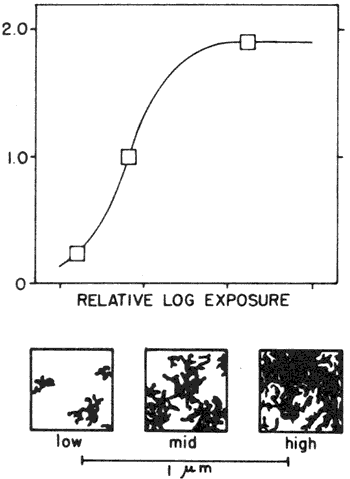
Figure 7. D log E curve of albumen paper showing schematized representations of the image deposit in low-, medium-, and high-density areas.
An important aspect of image formation in all printing-out papers is the increase in both particle size and number with increasing exposure. At low densities, the size of the image particles is at a minimum and their degree of dispersity is at a maximum. Because of their extreme smallness, at low densities the image particles are difficult to resolve in the electron micrographs. However, the typical appearance of albumen print images at various densities is shown schematically in Fig. 7. These schematic representations of the image deposit approximately correlate to the three density levels indicated on the D log E curve above.
Comparison with Developing-Out Print Materials
Figure 8. D log E curve of typical developing-out paper showing schematized representations of the image deposit in low-, medium-, and high-density areas.
Figure 9. Cross-section electron micrograph from high-density area of experimental albumen print toned in a gold chloride/sodium carbonate toner.
It is useful to compare the albumen print image structure shown in Fig. 7 with a schematized version (at the same scale) of the image structure of a typical developing-out paper, shown in Fig. 8. There is a large size differential between individual image particles in the two types of paper. Developing-out papers typically contain much larger silver halide crystals than do albumen prints. In images produced by direct development, the action of the developer converts the entire silver halide crystals to metallic silver in the form of a filament bundle. This imparts a relatively large minimum size to the basic structural unit of developed images, even at very low densities. The development mechanism also ensures that the filaments are closely packed spatially. These features give developed images distinct advantages over printed-out images in resisting attack by oxidants, especially in low-density areas.
The relatively greater resistance to deterioration of highlight areas of developed-out prints (compared to printed-out images) may be seen in the data from an incubation experiment comparing both types. The data in Table I are from a typical experiment of this kind, in which laboratory-made albumen prints and prints made on a contact-speed develop-out paper (Kodak Azo paper, grade 2) were incubated together at 50°C, 86% RH, for 60 days. Shown in Table I are percent density changes from starting densities of 0.30, 0.50, 1.00, and 1.50 for each material. The filters used for the red and blue reflection density measurements were Kodak Wratten filters 25 and 47B, respectively. The albumen prints were made by using two coatings of albumen (containing 1.25% NH4Cl by weight), sensitized with a 10% AgNO3/1% citric acid solution, exposed to a 1000 W mercury vapor lamp (NuArc GW 114), toned with a gold chloride/borax toner, and fixed in 10% sodium thiosulfate. The developed-out prints were made on Kodak Azo paper, grade 2, developed in Kodak developer D-72 1:1, and fixed in Kodak rapid fixer. Before the density changes in Table I were calculated, stain density (measured in nonimage areas) was subtracted from the image densities to correct for differing stains. The albumen prints had much greater percentage losses at the 0.30, 0.50, and 1.00 densities than the developed-out prints did. Similar results were obtained when other types of printing-out papers were compared with a bromide enlarging paper. The small particle size and high degree of dispersity characteristic of highlight areas of printing-out papers are the structural features responsible for their inferior image stability.
Effects of Gold Toning on Image Structure
Historically, nearly all albumen prints were gold toned prior to fixation. Numerous toner formulae were used, with various consequences for image color and stability. In our incubation experiments, gold thiocyanate toners provided more image stability in albumen prints than did alkaline gold toners containing sodium carbonate or sodium acetate. On the microstructural level, gold toning causes various degrees of distortion of the original particle morphology. Depending on the formula used, after toning such general features as the presence or absence of a crust, the grouping of particles into colonies, and the depth of image material in the coating are the same, but the Particle size decreases and individual particles are more irregular and elongated. A few become almost circular, with one or two thickened areas. Some gold toners tried in this study produced large changes in particle morphology in albumen prints, whereas others produced more subtle changes. Figure 9 shows an albumen print image showing considerable distortion of the original spherical particle shape due to toning with a gold chloride/sodium carbonate toner. The elongated, circular shape seen in some particles here is even more prominent in printed-out images which have been toned with platinum, a technique used mainly with matte collodion printing-out papers.
The toning process substitutes gold for some of the silver in the image.8-9 The final albumen print image contains 20-25% gold.10 Gold(I) ions will oxidize silver atoms on a one-for-one basis and take their place in the silver lattice. This is the likely cause for the observed distortion of particle shape during toning, and it provides evidence against the view of gold toning as merely a superficial deposition of gold. Gold(III) ions, if present, will oxidize three silver atoms for each gold atom reduced. The tendency for gold toning to diminish the particle size maybe rationalized through the oxidative nature of the toning process and the presence of some trivalent gold. The starting point for all the common toner formulae was gold chloride (chlorauric acid, HAuC14.3H20), a gold(III) complex.
Microstructural Changes During Deterioration
Figure 10. Area comparable to that of Fig. 9 after 48 days of incubation at 50°C, 95% RH.
For the studies of image deterioration in albumen prints, both 19th-century and laboratory-made prints were used. The view of the deterioration process that emerged is one in which the image is wholly re-formed through a continuing cycle of oxidation and reduction. The extreme small size of the individual image particles (with their consequent enormous surface area relative to their mass) renders them thermodynamically unstable, Gold toning may retard but does not fundamentally change this behavior. Albumen prints displayed rapid and dramatic changes in image microstructure under attack by moist air at moderate temperature. These microstructural changes are so extensive that they cannot be rationalized except through an oxidative-reductive mechanism. Structural features such as particle size, number, and spatial distribution help determine the degree of resistance of a given image toward oxidative attack. These features are important not only because they affect the oxidation step of the cycle, but also for their influence on the re-reduction of the silver ions formed when the original image was oxidized.
Figure 11. Size frequency distribution of image particles inside marked areas of Figs. 9 and 10; experimental albumen print. (—) Before incubation, 5.5-nm particles, 649 total particles. (- - -) After incubation, 7.5-nm particles, 195 total particles.
Initial Stages of Image Deterioration. The microstructural changes which occur in albumen prints incubated at 50°C, 95% RH illustrate how the structural factors of particle size, size frequency distribution, and spatial distribution are related. Figure 9 shows a micrograph of a high-density area near the upper surface of a newly made albumen print. A comparable area after 48 days of incubation is shown in Fig. 10. The particles inside the marked boxes were counted and measured with a reticle magnifier. Figure 11 shows the particle size frequency distribution before and after incubation; the mean particle diameters (measured on longest axis) and the total particle counts before and after incubation are given in the caption.
The image structure before incubation (Fig. 9) is characterized by a small mean particle diameter (5.5 nm), a wide range of particle diameters, and an even, relatively close spatial distribution. After only four days of incubation (not shown), many of the smallest particles had disappeared, and the mean particle diameter had increased; Weyde11 reported similar changes for small silver particles in gelatin. The particles were much rounder, losing the elongated and irregular outline induced by gold toning. These initial changes in microstructure have probably occurred to some degree in all surviving 19th-century albumen prints.
After 48 days of incubation (Fig. 10), complete re-formation of the image is evident. The mean particle size has increased considerably. and the total number of particles has sharply
decreased. These changes are typical of high-density areas of albumen prints during initial phases of deterioration. Figure 12 shows this initial re-formation in schematic form. At this point in the deterioration, the visual appearance of the albumen print image has changed considerably; low-density areas have faded to invisibility, while middletone and high-density areas have both lost density and become more reddish.
When a silver nitrate/citric acid sensitizing solution is used and the image particles are grouped together in colonies, as in Fig. 4, the process of re-formation is similar, but the colonies tend to retain their general shape and location in the coating. The grouping of particles in this way is advantageous in resisting oxidative attack, and in incubation studies coatings like those of Fig. 4 proved to be more stable than ones with a more even spatial distribution. The increased stability was particularly evident in highlight areas.
Predicting Microstructural Changes
Figure 12. Schematized representation of initial stages of deterioration in albumen prints, showing a decrease in the number of particles and an increase in average particle diameter.
What microstructural changes are likely to occur depend on the initial configuration of the image. Assuming a constant level of oxidant concentration and restricting the discussion to images with particle diameters of 50 nm or less, there are three operative factors: particle size, size frequency distribution, and spatial distribution. Size is important because smaller particles have a faster effective rate of attack. If a range of sizes is present, then smaller particles will disappear before larger ones. Image deterioration is a process which includes both oxidation and reduction; while small size favors oxidation, larger particles appear to act as favorable sites for the reduction of mobile silver ions formed during the oxidation step. This is why the initial stages of deterioration are characterized by a narrowing of the range of particle diameters and a decrease in the total number of particles. Although new particles are constantly being nucleated, they have little chance to grow at this stage because they cannot compete effectively with existing large particles for the silver ions necessary for growth.
Spatial distribution is important in two ways. When an individual particle is attacked by an oxidant, the silver ions and soluble silver compounds formed tend to diffuse and migrate away from it in all directions. Close packing of particles favors reduction of the silver ions because it tends to keep the silver ion concentration high. Close packing also increases the likelihood of reduction by providing other particles nearby which could serve as preferential sites for reduction. The silver ions from an isolated single particle are not as likely to encounter circumstances favorable for reduction back to metallic silver.
From the above discussion and the schematized image structure shown in Fig. 3, the extremely poor image stability of highlight areas of printing-out papers can be readily understood. In low-density areas the conditions are most favorable for the oxidative step and least favorable for reduction of the resulting silver ions. The micrographs of low-density areas showed a gradual withering away of image material rather than the re-formation which takes place in high-density areas.
Figure 13. Cross-section electron micrograph from high-density area of a 19th-century albumen print in average condition.
Figure 14. Area comparable to that of Fig. 13 after 90 days incubation at 50°C, 95% RH.
Figure 15. Size frequency distribution of image particles inside marked areas of Figs. 13 and 14, 19th-century albumen print. (—) Before incubation, 8.1-nm particles, 202 total particles. (- - -) After incubation, 6.8-nm particles, 343 total particles.
Figure 16. Schematized representation of later stages of deterioration in albumen prints showing a tendency for the image to re-form into smaller, more numerous, and more evenly spaced particles.
The microstructural changes typical of later stages of image deterioration in albumen prints are notably different from earlier stages. They can be observed when a 19th-century albumen print (in which some deterioration has already taken place) is subjected to high-humidity incubation. The visual consequences of these microstructural changes are continuing density loss and a shift to even yellower and redder image hues. Figure 13 is a micrograph from a high-density area of 19th-century albumen print in average condition. The changes produced in a comparable area of the coating after 90 days of incubation at 50°C, 95% RH are shown in Fig. 14. Figure 15 shows the size frequency distribution before and after incubation; the particle counts and mean particle diameters are given in the caption. These micrographs illustrate the second characteristic type of microstructural change observed in this study: a tendency for the image to re-form into smaller, more numerous, and evenly spaced particles. This is shown schematically in Fig. 16.
The fact that smaller and more numerous particles are formed at this stage seems almost contradictory to what occurred in the early stages of deterioration. Actually the same forces are at work, but the dominant factor is no longer the competitive advantages of existing larger particles because all the particles are close to the same size. From this point on, the range of particle diameters will continue to be narrow, and spatial distribution will be the key factor. The changes which occur in the later stages of deterioration can best be understood as moving toward an equilibrium condition where all the particles are of uniform size and are uniformly spaced. New particles will nucleate and grow in any empty area far enough removed from an existing particle to negate its advantage as a preferential site for reduction. Feldman12 reported the nucleation and growth of colloidal silver particles [in?] the empty gelatin surrounding bundles of filamentary silver undergoing oxidative attack.
The particle size at equilibrium will depend on the total amount of silver and silver ion available to take part in the continuing cycle of oxidation and reduction. Filling the empty areas decreases the mean particle size, but as deterioration progresses the particle size will further decrease because of the migration of silver ions away from the area of the main image deposit. Some silver ions are drained away to form a crust of closely spaced particles at the uppermost surface, while others migrate into lower areas of the coating.
The image structure of albumen prints is determined by the photolytic mechanism of image formation. Structural features typical of photolytic images are responsible for the inferior image stability of albumen prints, in particular the rapid loss of highlight detail. The fact that in incubation experiments moist air produced dramatic transformations in albumen print image microstructure demonstrates the vulnerability of albumen prints to oxidative-reductive deterioration and underscores the need to control the relative humidity of the storage environment for these objects.
We acknowledge the support of the National Endowment for the Humanities Research Resources Program and the National Museum Act (administered by the Smithsonian Institution) for making this research possible. In addition, the assistance of Dr. Wesley T. Hanson and Eastman Kodak Company are gratefully acknowledged.
1. J. M. Reilly, D. Severson, and C. McCabe, Image Deterioration in Albumen Prints; In Preprints of Contributions, 9th International Congress of the International Institute for Conservation of Artistic and Historic Works, Washington, DC, September 1982, pp. 16-65.
2. Leo Baekeland, In The Photograph, H. Snowden Ward, Ed., Dawbarn and Ward, London, 1897, Vol. IV, pp. 269-271.
3. J. M. Eder and F. Wentzel, Die photographischen Kopirverfahren mit Silbersalzen (Positiv-Prozess), Wilhelm Knapp, Halle, 1928, Ch. 10.
4. A. Miethe, Lehrbuch der Praktischen Photographie, Wilhelm Knapp, Halle, 1902, Ch. 5.
5. H. Stiefel, Sensitized Papers, How Made and Used, The Adams Press, New York, 1894, Ch. 10.
6. K. B. Hendriks, The Archivist 10: 6(1983).
7. B. Fergg, Z. Wiss. Photogr. Photophys. Photochem. 52: 24(1957).
8. F. Formstecher, Camera (Luzern) 3; 193(1924).
9. A. W. Henn and B. 0. Mack, Photogr. Sci. Eng. 9: 378(1965).
10. F. Novak, In Jahrbuch fur Photographie und Reproductionstechnik 1902, J. M. Eder, Ed., Wilhelm Knapp, Halle, 1902, pp. 183-185.
11. E. Weyde, Photogr. Sci. Eng. 16: 283(1972).
12. L. Feldman, J. Appl. Photogr. Eng. 7: 1(1981).